
两种蔷薇珊瑚骨骼微结构观察及钙化过程分析
作者简介:耿涛年(1991—), 男, 在读硕士生。
收稿日期: 2016-02-15
要求修回日期: 2016-09-08
网络出版日期: 2017-01-19
基金资助
国家自然科学基金项目(31460555)
国家海洋局项目(HZ2012-174)
“中西部高校提升综合实力”资助项目
Skeletal microstructure observations and calcification process analysis of two species of Montipora
Received date: 2016-02-15
Request revised date: 2016-09-08
Online published: 2017-01-19
Supported by
National Natural Science Foundation of China (31460555)
State Oceanic Administration Project (HZ2012-174)
“Central and Western Colleges and Universities Enhance the Comprehensive Strength” Funding Projects
Copyright
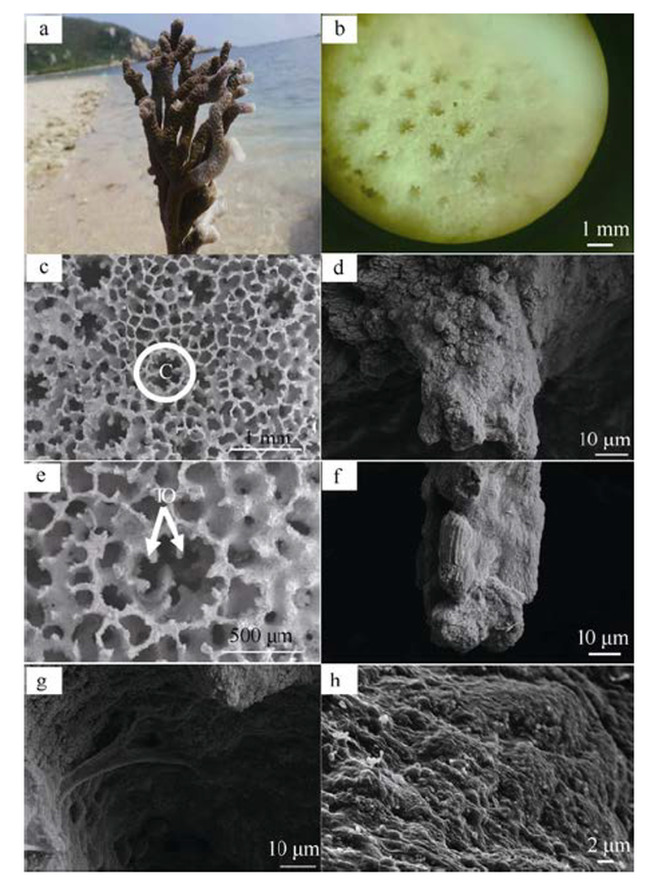
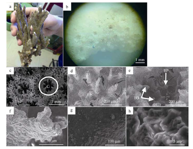
蔷薇珊瑚属是印度—太平洋地区第二大造礁石珊瑚属, 本研究选取两种分枝状蔷薇珊瑚——指状蔷薇珊瑚Montipora digitata与脆蔷薇珊瑚Montipora fragilis, 利用扫描电子显微镜和能谱仪观察分析其骨骼微结构和钙化过程。结果表明, 两种蔷薇珊瑚骨骼基本组成结构(包括珊瑚杯、隔片、体壁、鳞板等)大体相同, 但也存在诸多细节差异。指状蔷薇珊瑚共骨为平滑型网状, 表面有较短的小刺(长约40μm), 珊瑚杯之间间隔较大(约1mm), 第一轮隔片除直接隔片大而呈薄片状外, 其他隔片发育不完整, 隔片刺较发达呈扁平狭板状。脆蔷薇珊瑚共骨为瘤突型网状, 表面有众多的小刺(长约100μm), 珊瑚杯之间有隆起的脊, 第一轮隔片退化成短刺状。脆蔷薇珊瑚体壁上的小孔比指状蔷薇珊瑚的大。蔷薇珊瑚骨骼主要成分为文石晶体, 多为簇状, 少数为鹅卵石状。能谱分析表明, 珊瑚碳酸钙骨骼并非直接生成, 而是经历以下4个过程: 1)钙的富集, 形成钙点; 2)钙结合碳与氧, 此时碳多氧少; 3)文石晶体趋于规则, 碳元素减少, 氧元素增多; 4)大量的文石晶体聚集成羽簇, 有序地排列形成成熟的骨骼。
耿涛年 , 姚雪梅 , 张颖 , 谢夏岭 , 崔敏 , 林道明 . 两种蔷薇珊瑚骨骼微结构观察及钙化过程分析[J]. 热带海洋学报, 2017 , 36(1) : 56 -64 . DOI: 10.11978/2016015
Montipora is the second species-rich scleractinian genus that is widely distributed in the Indo-Pacific Ocean. Montipora digitata and Montipora fragilis, two species of branching Montipora, from the South China Sea were selected in this study, and its skeletal microstructures and calcification processes were observed and analyzed using Scanning Electron Microscopy (SEM) combined with Energy Dispersive Spectrometer (EDS). The results showed that the two species of Montipora skeletal basic elements were substantially the same (including calice, septum, theca, dissepiment, etc.), but there were many differences in details. Coenosteum surface of M. digitata was glabrous and recticular formation with shorter spines (about 40 μm) and each calices (about 1 mm) were separated by big interval. The septa of the first cycle was poorly developed except that the shape of its direct septum was sheet. In addition, teeth along the margin of septa developed well and had flat shape. The coenosteum surface of M. fragilis was tuberculate and recticular formation with many small spines (about 100 μm) and irregular nodular ridge between the calices. Compared with M. digitata, the first-cycle septa of M. fragilis had degenerated teeth. Main ingredients of the two kinds of coral skeletons were aragonite crystals, with most being tufted-crystals and a few being pebble-crystals. EDS analysis showed that the components of calcium carbonate from the skeleton were not directly generated, but formed in the following four processes: 1) the calcium was concentrated to develop calcium point; 2) calcium combined with carbon and oxygen, and the content of carbon was higher than that of oxygen; 3) aragonite crystals tended to look regular, and the content of carbon decreased and that of oxygen increased; and 4) a lot of aragonite crystals gathered into sclerodermite in orderly arrangement to form mature skeleton.
Fig. 1 Skeletal basic structure unit of M. digitata图1 指状蔷薇珊瑚骨骼基本结构单元 |
Fig. 2 Skeletal basic structure unit of M. fragilis图2 脆蔷薇珊瑚骨骼基本结构单元 |
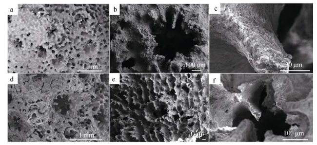
Fig. 3 Skeletal shadow branch surface structures of Montipora digitata and Montipora fragilis图3 指状蔷薇珊瑚与脆蔷薇珊瑚骨骼背阴面表面结构 |
Fig. 4 Skeletal internal structure characteristics of M. digitata and M. fragilis图4 指状蔷薇珊瑚与脆蔷薇珊瑚骨骼内部结构特征 |
Fig. 5 Microstructure of M. fragilis growing point图5 脆蔷薇珊瑚的生长点表观结构 |
Fig. 6 Section of M. fragilis growing point图6 脆蔷薇珊瑚生长点纵切 |
Fig. 7 Montipora skeletal calcification process图7 蔷薇珊瑚骨骼钙化过程 |
Tab. 1 Weight and atomic percentage of each element in calcification process of skeleton表1 蔷薇珊瑚骨骼钙化过程各元素重量百分比和原子百分比 |
| 元素 | 重量比 | 原子比 | ||||||
|---|---|---|---|---|---|---|---|---|
| A | B | C | D | A | B | C | D | |
| C | 0 | 34.61 | 20.10 | 18.13 | 0 | 57.83 | 36.32 | 28.30 |
| O | 0 | 17.10 | 30.09 | 49.89 | 0 | 21.46 | 40.81 | 58.45 |
| Ca | 79.05 | 38.16 | 38.76 | 26.65 | 92.28 | 19.11 | 20.98 | 12.47 |
| I | 20.95 | 10.12 | 11.06 | 5.33 | 7.72 | 1.60 | 1.89 | 0.79 |
注: A为最初钙沉积点(生长点纵切); B为钙点扩大化(分枝中上部纵切); C为钙点平铺(分枝中下部纵切); D为形成羽簇(分枝底部纵切) |
The authors have declared that no competing interests exist.
| [1] |
|
| [2] |
|
| [3] |
|
| [4] |
|
| [5] |
|
| [6] |
|
| [7] |
|
| [8] |
|
| [9] |
|
| [10] |
|
| [11] |
|
| [12] |
|
| [13] |
|
| [14] |
|
| [15] |
|
| [16] |
|
| [17] |
|
| [18] |
|
| [19] |
|
| [20] |
|
| [21] |
|
| [22] |
|
| [23] |
|
| [24] |
|
| [25] |
|
| [26] |
|
| [27] |
|
| [28] |
|
| [29] |
|
| [30] |
|
| [31] |
|
| [32] |
|
| [33] |
|
| [34] |
|
/
| 〈 |
|
〉 |


