
海参抗菌肽的活性筛选与功效评价
|
袁华标(1997—), 男, 广东湛江人, 硕士研究生, 从事海洋生物活性肽的分离纯化与活性研究。email: |
Copy editor: 姚衍桃
收稿日期: 2022-09-30
修回日期: 2022-11-09
网络出版日期: 2022-12-05
基金资助
广东省海洋经济发展专项(粤自然资合[2022]036)
广东省海洋经济发展专项(粤自然资合[2020]036)
广东省海洋经济发展专项(粤自然资合[2020]041)
广东省自然科学基金项目(2022A1515010767)
广东省重点领域研发计划(2020B1111030004)
中国科学院南海生态环境工程创新研究院自主部署项目(ISEE2021PY05)
海南省重点研发计划(ZDYF2021SHFZ109)
Screening and efficacy evaluation of antibacterial peptides from Holothuroidea
Copy editor: YAO Yantao
Received date: 2022-09-30
Revised date: 2022-11-09
Online published: 2022-12-05
Supported by
Marine Economy Development Special Project of Guangdong Province(GDNRC[2022]036)
Marine Economy Development Special Project of Guangdong Province(GDNRC[2020]036)
Marine Economy Development Special Project of Guangdong Province(GDNRC[2020]041)
National Natural Science Foundation of Guangdong, China(2022A1515010767)
Key-Area Research and Development Program of Guangdong Province(2020B1111030004)
Institution of South China Sea Ecology and Environmental Engineering, Chinese Academy of Sciences(ISEE2021PY05)
Key Research and Development Program of Hainan Province(ZDYF2021SHFZ109)
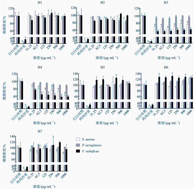
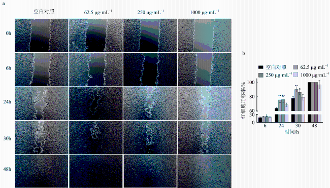
本文将已有文献报道的海洋动物源抗菌肽(毒素除外)与海参蛋白组进行比对, 结合生物信息学软件, 设计、筛选海参抗菌肽, 并对其理化特性、抗菌活性与机制、生物相容性与细胞活性等进行研究。研究结果发现, 通过软件模拟与抗菌活性预测, 最终筛选并固相合成7条海参抗菌肽(H1—H7)。其中, H4(RVHRFLRR)可通过细菌膜电位去极化、膜透化等方式, 抑制金黄色葡萄球菌(Staphylococcus aureus)、铜绿假单胞菌(Pseudomonas aeruginosa)、创伤弧菌(Vibrio vulnificus)生长, 在较低浓度(31.25µg·mL-1)时, 可使S. aureus细菌总数降至84.93%±4.21%(p<0.01), P. aeruginosa细菌总数降至95.92%±0.52%(p<0.01), V. vulnificus细菌总数降至77.14%±1.37%(p<0.01)。H4生物相容性好, 在<1000µg·mL-1时均未表现出溶血性或细胞毒性, 其可能通过与细胞膜表面受体(EGFR、VEGFR2、FGFR1等)发生相互作用进而促进细胞迁移。在浓度为62.50µg·mL-1和250µg·mL-1时, H4均可显著促进L929细胞发生迁移(p<0.05)。实验结果表明, 海参抗菌肽H4生物相容性好, 兼有抗菌、促愈合功效, 可望作为一种潜在功能物质用于感染创面的修复与再生。
袁华标 , 黄靖彤 , 万鹏 , 蔡冰娜 , 潘剑宇 , 张煜航 , 凌娟 , 陈华 . 海参抗菌肽的活性筛选与功效评价[J]. 热带海洋学报, 2023 , 42(4) : 184 -194 . DOI: 10.11978/2022208
In this paper, we compared the existing marine animal origin antibacterial peptides reported (Toxin except) with the holothurian proteome, combined with bioinformatics software to design, screen holothurian antibacterial peptides, and to investigate their physicochemical properties, antibacterial activity, mechanism, biocompatibility and cellular activity. It was found that through software simulation and antibacterial activity prediction, 7 holothurian bacteriostatic peptides (H1~H7) were finally screened and solid-phase synthesized. Among them, H4 (RVHRFLRR) could inhibit the growth of Staphylococcus aureus, Pseudomonas aeruginosa, Vibrio vulnificus through bacterial membrane potential depolarisation or membrane permeabilisation. At a lower concentration (31.25 µg·mL-1), it reduced the total bacterial count of S. aureus to 84.93%±4.21%(p<0.01), P. aeruginosa to 95.92%±0.52%(p<0.01) and V. vulnificus to 77.14%±1.37%(p<0.01). It was also found that H4 was biocompatible and did not exhibit haemolytic or cytotoxic properties at < 1000 µg·mL-1. In addition, H4 may interact with cell membrane receptors (EGFR, VEGFR2, FGFR1, etc.) to promote cell migration. H4 significantly promoted the migration of L929 cells at both 62.50 µg·mL-1 and 250 µg·mL-1 (p<0.05). The above experimental results indicate that holothurian antibacterial peptide H4 is biocompatible, has both antibacterial and pro-healing effects, and can be used as a potential functional active substance for the repair and regeneration of infected wounds.
Key words: Holothuroidea; antibacterial peptide; bioinformatics; cell migration
表1 肽序列抗菌活性预测Tab. 1 Prediction of peptide sequences for bacteriostatic activity |
| 序号 | 序列 | SVM | RF | ANN | DA | 蛋白来源 |
|---|---|---|---|---|---|---|
| 1 | RVHRFLRR | 1.00 | 0.61 | AMP | 0.89 | 组蛋白H2A |
| 2 | KKPSKKPATKKAAKKKPAAKPKKAVKPKKPAAKKPAKKTSPKKKTAKKPAKKA | 1.00 | 0.68 | AMP | 0.99 | 组蛋白H1β |
| 3 | PGKKTPQKKKPAAKKTKKPV | 1.00 | 0.67 | AMP | 0.99 | 组蛋白H5 |
| 4 | AKKPVRRLLQRRRRLRRRER | 1.00 | 0.67 | AMP | 0.98 | 组蛋白H5 |
| 5 | KPKKAVKPKKP | 1.00 | 0.62 | AMP | 0.97 | 组蛋白H1β |
| 6 | KKAAKKKPAAKPKKAVKPKKP | 1.00 | 0.69 | AMP | 1.00 | 组蛋白H1β |
| 7 | KKAAKKKPAAKPKKAVKPKKPAAKKPAKK | 1.00 | 0.71 | AMP | 1.00 | 组蛋白H1β |
| 8 | KPAIRRLARR | 1.00 | 0.62 | AMP | 0.77 | 组蛋白H4 |
| 9 | RSARAGLQFPVGRVHRFLRR | 0.99 | 0.81 | AMP | 0.98 | 组蛋白H2A |
| 10 | RGKGGKAWAKAKSRSARAGLQFPVGRVHRFLRR | 0.99 | 0.92 | AMP | 1.00 | 组蛋白H2A |
| 11 | KKAAKKK | 0.98 | 0.57 | AMP | 0.53 | 组蛋白H1β |
| 12 | FLGIKQTLKSLRQGKAKLII | 0.98 | 1.00 | AMP | 1.00 | 核糖体L30 |
| 13 | KAWAKAKSRSARAGLQFPVGRVHRFLRR | 0.96 | 0.97 | AMP | 1.00 | 组蛋白H2A |
| 14 | KKKPVAAKKPVRRLLQRRRR | 0.95 | 0.71 | AMP | 1.00 | 组蛋白H5 |
| 15 | ILRLAVGLKGLPSVNPAWLV | 0.93 | 0.97 | AMP | 0.99 | 组蛋白H3 |
| 16 | ANFITHPYKRILRLAVGLKG | 0.92 | 0.98 | AMP | 0.99 | 组蛋白H3 |
| 17 | IKVELKRLAANNVLVHTKGT | 0.90 | 0.86 | AMP | 0.84 | 组蛋白H5 |
| 18 | IFVKTLTGKT | 0.81 | 0.40 | AMP | 0.82 | 海参泛素 |
| 19 | KYFLGIKQTLKSLRQGKAKLIIL | 0.81 | 0.95 | AMP | 0.99 | 核糖体L30 |
| 20 | RGKGGKAWAKAK | 0.74 | 0.47 | AMP | 0.41 | 组蛋白H2A |
| 21 | MQIFVKTLTGKTITL | 0.38 | 0.23 | NAMP | 0.25 | 海参泛素 |
| 22 | MTGRGKGGKAWAKAKSRSARAGLQFPVGRVHRFLRRGNY | 0.36 | 0.51 | AMP | 0.92 | 组蛋白H2A |
| 23 | FDKGKLKTVETQEKNTLPTKDTIDQEK | 0.21 | 0.11 | NAMP | 0.03 | 胸腺素 |
| 24 | MQIFVKTLTGKTITLEVEPSDTIENVKSKIQDKEGIPPDQQRLIFAGKQLEDGRTLSDYNIQKESTLHLVLRLR | 0.21 | 0.26 | NAMP | 0.04 | 海参泛素 |
| 25 | MSDKPDVSEVTK | 0.06 | 0.07 | NAMP | 0.01 | 胸腺素 |
| 26 | MSDKPDVSEVTKFDKGKLKTVETQEKNTLPTKDTIDQEKAAS | 0.02 | 0.04 | NAMP | 0.00 | 胸腺素 |
| 27 | STLHLVLRLR | 0.00 | 0.33 | NAMP | 0.15 | 海参泛素 |
注: AMP表示抗菌肽, NAMP表示非抗菌肽; SVM>0.50即为AMP, RF>0.50即为AMP, DA>0.50即为AMP。表中加粗字体表示挑选出的7条抗菌肽序列 |
表2 H1—H7理化性质分析Tab. 2 Analysis of the physical and chemical properties of H1~H7 |
| 抗菌肽 | 肽序列 | 疏水性/% | 净电荷/C | pI | 分子量/Da |
|---|---|---|---|---|---|
| H1 | RGKGGKAWAKAK | 33.30 | +5.00 | 11.83 | 1257.51 |
| H2 | RSARAGLQFPVGRVHRFLRR | 45.00 | +11.10 | 13.18 | 2379.81 |
| H3 | KAWAKAKSRSARAGLQFPVGRVHRFLRR | 46.43 | +9.10 | 13.19 | 3250.86 |
| H4 | RVHRFLRR | 37.50 | +4.10 | 12.96 | 1139.38 |
| H5 | KPKKAVKPKKP | 45.45 | +6.00 | 11.21 | 1248.63 |
| H6 | KKAAKKK | 28.57 | +5.00 | 11.11 | 801.05 |
| H7 | KKAAKKKPAAKPKKAVKPKKP | 47.62 | +11.00 | 11.51 | 2270.93 |
图3 H4与L929细胞共孵育24h后的荧光染色照片(a)、死细胞数量计数(b)和活细胞数量计数(c), 以及H4溶血性测定(d)与空白对照组比较, *表示p<0.05, **表示p<0.01 Fig. 3 Photograph of live-dead cell staining (a), number of live cells (b), number of dead cells (c) and hemolytic properties of H4 (d). Compared with control group, * stands for p<0.05, and ** stands for p<0.01 |
图4 H4与L929细胞共孵育24h后光镜(100×)照片(a)和细胞存活率(b)Fig. 4 Results of CCK8 assay for cell proliferation viability. Photographs under 100x light microscope after 24 h incubation (a) and cell viability (b) |
| [1] |
侯晓艳, 2019. 基于生物信息学与多肽组学的花椒籽抗菌肽筛选及抑菌机理研究[D]. 成都: 四川农业大学.
|
| [2] |
李成华, 2007. 栉孔扇贝核心组蛋白的基因结构及H2A抗菌活性的研究[D]. 青岛: 中国科学院海洋研究所.
|
| [3] |
李冠楠, 夏雪娟, 隆耀航, 等, 2014. 抗菌肽的研究进展及其应用[J]. 动物营养学报, 26(1): 17-25.
|
| [4] |
马健, 2010. 海参再生研究进展[J]. 安徽农业科学, 38(11): 5694-5695, 5768.
|
| [5] |
聂竹兰, 李霞, 2006. 海参再生的研究[J]. 海洋科学, 30(5): 78-82.
|
| [6] |
张婷婷, 高珊, 矫建, 等, 2018. 香丹注射液溶血性实验及药物安全性检测问题探讨[J]. 中国药事, 32(4): 529-532.
|
| [7] |
朱宁艺, 2021. 抗菌肽Mastoparan-C新型类似物的设计、合成、构效关系及其抑制和逆转抗生素耐药性作用研究[D]. 兰州: 兰州大学.
|
| [8] |
|
| [9] |
|
| [10] |
|
| [11] |
|
| [12] |
|
| [13] |
|
| [14] |
|
| [15] |
|
| [16] |
|
| [17] |
|
| [18] |
|
| [19] |
|
| [20] |
|
| [21] |
|
| [22] |
|
| [23] |
|
| [24] |
|
| [25] |
|
| [26] |
|
| [27] |
|
| [28] |
|
| [29] |
|
| [30] |
|
| [31] |
|
| [32] |
|
| [33] |
|
/
| 〈 |
|
〉 |


