
热带海洋学报 ›› 2023, Vol. 42 ›› Issue (6): 137-149.doi: 10.11978/2023027CSTR: 32234.14.2023027
广西北部湾施氏獭蛤卵巢发育、卵子和卵黄发生的研究
吴韬1,2( ), 潘英1,2(), 连昌朋1,2, 刘一鸣1,2, 徐炳杰1,2, 王超奇1,2, 杨凌1,2
- 1.广西大学动物科学技术学院, 广西 南宁 530004
2.广西高校水生生物健康养殖与营养调控重点实验室, 广西 南宁 530004
-
收稿日期:2023-03-03修回日期:2023-05-05出版日期:2023-11-10发布日期:2023-04-25 -
作者简介:吴韬(1997—), 男, 广西南宁市人, 硕士研究生, 从事海洋贝类遗传育种研究。email: 214850470@qq.com
-
基金资助:广西创新驱动发展专项(AA19254032); 国家重点研发计划项目(2018YFD0901400)
The study on ovarian development, oogenesis and vitellogenesis of Lutraria sieboldii in Beibu Gulf of Guangxi
WU Tao1,2(), PAN Ying1,2(), LIAN Changpeng1,2, LIU Yiming1,2, XU Bingjie1,2, WANG Chaoqi1,2, YANG Ling1,2
- 1. College of Animal Science and Technology, Guangxi University, Nanning 530004, China
2. Key Laboratory of Aquatic Healthy Breeding and Nutrition Regulation of Guangxi Universities, Nanning 530004, China
-
Received:2023-03-03Revised:2023-05-05Online:2023-11-10Published:2023-04-25 -
Supported by:Guangxi Innovation Driven Development Project(AA19254032); National Key Research and Development Program of China(2018YFD0901400)
摘要:
为探究广西北部湾海域施氏獭蛤卵巢发育、卵子和卵黄发生, 采用组织切片分析卵巢周年变化, 并利用光学和电子显微镜观察雌性生殖细胞的变化过程。结果表明, 广西北部湾海域施氏獭蛤卵巢发育周期为一年, 每年12月至翌年4月性腺饱满, 为繁殖盛期, 产卵高峰期略滞后于性腺成熟期, 每期5%~15%个体发育滞后于群体。卵子发生过程中, 雌性生殖细胞逐渐脱离滤泡壁, 进入滤泡腔。卵子发育过程中, 卵径由6.9~8.3μm变大至70.0~74.9μm, 细胞质内细胞器数量增加, 出现卵黄粒, 自噬泡吞噬脂滴和线粒体。卵黄发生期间, 核仁经染色后分为两部分, 颜色深浅不一, 核质间出现物质交换, 卵母细胞质膜内出现多泡小体, 在靠近卵周隙处形成微吞饮泡, 分别从卵周隙和滤泡吸收外源性卵黄物质。此外, 本实验还观察到施氏獭蛤存在滤泡混合型和滤泡共存型雌雄同体, 性转换方向表现为雌性向雄性转换。本研究结果为施氏獭蛤的人工繁育提供基础资料。
引用本文
吴韬, 潘英, 连昌朋, 刘一鸣, 徐炳杰, 王超奇, 杨凌. 广西北部湾施氏獭蛤卵巢发育、卵子和卵黄发生的研究[J]. 热带海洋学报, 2023, 42(6): 137-149.
WU Tao, PAN Ying, LIAN Changpeng, LIU Yiming, XU Bingjie, WANG Chaoqi, YANG Ling. The study on ovarian development, oogenesis and vitellogenesis of Lutraria sieboldii in Beibu Gulf of Guangxi[J]. Journal of Tropical Oceanography, 2023, 42(6): 137-149.
表1
施氏獭蛤形态参数"
| 数值 | 壳长/mm | 壳高/mm | 壳宽/mm | 体质量/g |
|---|---|---|---|---|
| 平均值 | 94.12 | 46.03 | 32.61 | 98.89 |
| 最大值 | 116.80 | 57.74 | 43.83 | 199.96 |
| 最小值 | 80.12 | 39.53 | 24.72 | 55.30 |
表1
图1
雌性施氏獭蛤性腺指数与水温的周年变化"

图1

图2
雌性施氏獭蛤肥满度与水温的周年变化"
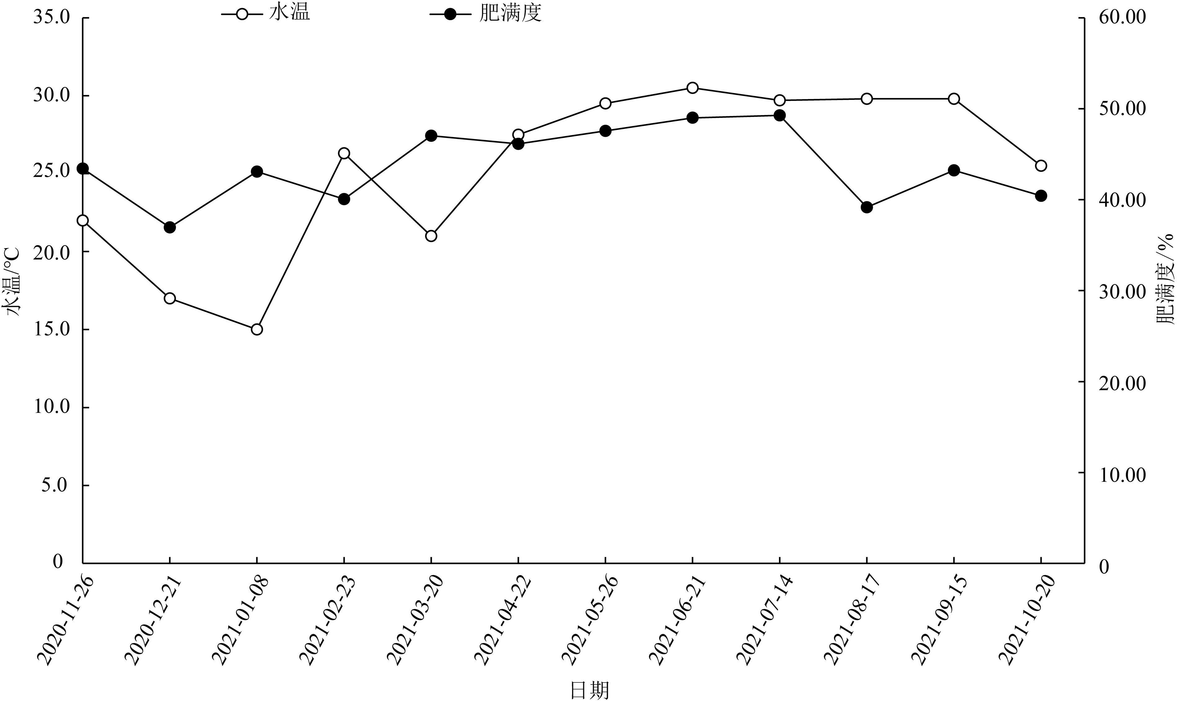
图2
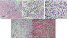
图3
施氏獭蛤各期卵巢的发育 a. 增殖期(Ⅰ); b. 生长期(Ⅱ); c. 成熟期(Ⅲ); d. 排放期(Ⅳ); e. 休止期(Ⅴ)"

图3
表2
施氏獭蛤各期卵巢的特点"
| 发育分期 | 各期特点 |
|---|---|
| 增殖期(Ⅰ) | 结缔组织包裹至软体部上缘, 性腺颜色透明, 雌雄外观无区别, 呈细线分布。结缔组织间形状不规则且狭长的滤泡构成性腺, 滤泡末端分支由少变多, 滤泡由小变大形成滤泡腔, 滤泡间隙较大。此时期滤泡腔大多为空腔, 滤泡壁厚, 滤泡内大部分为卵原细胞, 卵原细胞松散分布在滤泡壁上。此期为8月下旬—9月 |
| 生长期(Ⅱ) | 性腺增殖至软体部上缘, 仍无法从性腺颜色分辨雌雄。滤泡不断增大, 数量不断增多, 结缔组织减少, 滤泡间存在间隙。滤泡壁变薄, 滤泡壁上雌性生殖细胞数量增加, 部分次级卵母细胞生成卵柄。小部分成熟卵母细胞从滤泡壁脱落进入滤泡腔, 大量未发育成熟的卵母细胞占据着滤泡。此期为10月—11月下旬 |
| 成熟期(Ⅲ) | 性腺增殖至软体部腹缘和足基部, 呈白色, 肉眼可分辨雌雄。此期滤泡最大, 结缔组织退化, 滤泡相互挤压呈不规则形。滤泡壁变为单层, 其上残留少数初级卵母细胞。次级卵母细胞卵柄断裂进入滤泡腔内, 发育为成熟卵子, 滤泡腔内卵子形状不规则。此期为12月—次年3月底 |
| 排放期(Ⅳ) | 性腺仍饱满, 大部分成熟卵子排出体外, 腔内残留着少量成熟卵子与滤泡壁相连。滤泡壁变厚, 滤泡逐渐缩小, 呈现出大小不一的空腔, 滤泡间结缔组织开始增多。此期为次年4月 |
| 休止期(Ⅴ) | 软体部消瘦, 性腺变薄, 无法从软体部颜色分辨雌雄。滤泡间隙进一步加大, 结缔组织增长挤压滤泡。滤泡内已无成熟卵子, 滤泡壁上可见少量处于重吸收的卵原细胞, 滤泡壁变厚呈粗线条状。此期为5月下旬—8月中旬 |
表2
表3
广西北部湾施氏獭蛤卵巢发育各期的周年分布"
| 取样日期 | 盐度/‰ | 水温/℃ | 总数 | 雌雄同体数 | 卵巢发育分期 | ||||
|---|---|---|---|---|---|---|---|---|---|
| Ⅰ | Ⅱ | Ⅲ | Ⅳ | Ⅴ | |||||
| 2020-11-26 | 27.4 | 22.0 | 9 | 8 | 1 | ||||
| 2020-12-21 | 27.5 | 17.0 | 10 | 2 | 2 | 5 | 1 | ||
| 2021-01-08 | 29.4 | 15.0 | 9 | 1 | 8 | ||||
| 2021-02-23 | 30.9 | 26.3 | 14 | 4 | 10 | ||||
| 2021-03-20 | 30.0 | 21.0 | 11 | 3 | 8 | ||||
| 2021-04-22 | 30.0 | 27.5 | 12 | 10 | 2 | ||||
| 2021-05-26 | 28.0 | 29.5 | 12 | 12 | |||||
| 2021-06-21 | 28.4 | 30.5 | 7 | 1 | 1 | 1 | 5 | ||
| 2021-07-14 | 29.0 | 29.7 | 11 | 2 | 4 | 6 | |||
| 2021-08-17 | 30.0 | 29.8 | 10 | 1 | 1 | 1 | 1 | 3 | 4 |
| 2021-09-15 | 31.0 | 29.8 | 9 | 1 | 4 | 1 | 1 | 3 | |
| 2021-10-20 | 29.0 | 25.5 | 9 | 2 | 5 | 2 | |||
表3
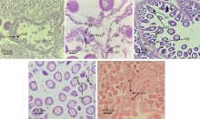
图4
施氏獭蛤卵子发生各期组织切片 a. 增殖期, 示卵原细胞(OG)、核仁(NU); b. 小生长期, 示卵原细胞(OG)、初级卵母细胞(PO)、核仁(NU)、滤泡细胞(FC); c. 大生长期, 示次级卵母细胞(SO)、核仁(NU)、卵原细胞(OG)、初级卵母细胞(PO); d.成熟期, 示成熟卵母细胞(OV)、核仁(NU)、卵原细胞(OG); e. 卵巢排放期, 示成熟卵子退化(DO)、核仁(NU)"

图4
表4
施氏獭蛤卵子发生各期特点"
| 发生分期 | 各期发生特点 |
|---|---|
| 增殖期 | 出现在Ⅰ—Ⅳ期卵巢。体积小、染色浅的卵原细胞大量增殖, 与滤泡细胞一起平行排列于滤泡壁基膜上 |
| 小生长期 | 出现在Ⅰ—Ⅳ期卵巢。卵原细胞体积增大, 分化形成生长较缓慢的初级卵母细胞, 细胞的大部分仍紧贴滤泡壁基膜, 细胞核内有一核仁常偏位, 细胞周围围绕着一层滤泡细胞。细胞内较为空虚, 染色质数量较少且不清晰, 细胞核染色较浅 |
| 大生长期 | 初级卵母细胞迅速生长, 细胞核内卵黄粒大量形成, 细胞质内细胞器大量形成, 生长期可延续到第一次减数分裂形成次级卵母细胞前, 出现在Ⅰ—Ⅳ期卵巢 |
| 成熟期 | 出现在施氏獭蛤卵巢发育的Ⅲ—Ⅳ期。此期成熟卵子体积达到最大, 成熟卵子呈不规则圆形。初级卵母细胞经过小、大生长期, 胞质内物质大量累积, 发育到次级卵母细胞, 逐渐脱离滤泡壁基膜, 后期卵柄断裂萎缩, 卵母细胞发育成熟进入滤泡腔内, 少数成熟卵子退化被滤泡重吸收 |
表4
图5
施氏獭蛤雌性生殖细胞发育各期超微结构 a. 卵原细胞(OG)和初级卵母细胞(PO), ×1000; b. 卵巢局部及次级卵母细胞(SO), ×1500; c. 成熟卵母细胞(OV), ×1500"

图5
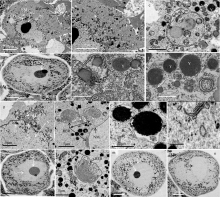
图6
施氏獭蛤卵子发生及卵黄发生的超微结构 a. 卵黄合成前期卵母细胞, 示细胞核(N)、核仁(NU)、核膜(NM)、染色质(CH)、滤泡(F)、基膜(BM)、卵柄(ES)、多泡小体(MB), ×1000; b. 卵黄合成前期, 示线粒体(M)、滑面内质网(SER)、脂滴(L)、滤泡(F)、微吞饮泡(MP), ×2000; c. 卵黄合成前期吞噬泡, 示线粒体(M)、卵黄(Y)、脂滴(L), ×5000; d. 卵黄合成期卵母细胞, 示细胞核(NU)、基膜(BM), ×300; e. 卵黄合成期, 示线粒体(M)、卵黄(Y)、脂滴(L)、核糖体(R), ×5000; f. 卵黄合成期, 示溶酶体(LY)、卵黄(Y)、线粒体(M), ×5000; g. 卵黄合成期细胞核与细胞质进行物质交换, 示染色质(CH)、细胞核(N)、核孔(NP), ×2000; h. 卵黄合成期, 示粗面内质网(RER)、核膜(NM)、卵黄(Y)、线粒体(M), ×2000; i. 卵黄合成期卵黄形成, ×5000; j. 卵黄合成期, 示高尔基复合体(G)、核糖体(R), ×2000; k. 卵黄合成后期卵母细胞, 示细胞核(N)、核仁(NU), ×300; l. 卵黄合成后期, 示粗面内质网(RER)、线粒体(M), ×2000; m. 成熟卵母细胞, 示细胞核(N)、核仁(NU), ×300; n. 成熟卵母细胞, 示细胞核(N), ×300"

图6
表5
施氏獭蛤内源性卵黄形成过程各细胞器特点"
| 细胞器 | 细胞器变化情况 |
|---|---|
| 线粒体 | 线粒体为细胞内部活动提供能量, 卵黄合成前期胞质中线粒体数量较少, 内部嵴少且清晰。随着卵黄的合成, 线粒体数量逐渐增多, 内部嵴模糊, 卵黄物质在线粒体基质中的内膜沉积并逐渐充满整个线粒体, 线粒体内膜逐渐溶解消失, 最终形成单层膜结构的卵黄粒( |
| 粗面内质网 | 卵黄合成期粗面内质网呈环形状和片条状, 片条状粗面内质网两端形成囊泡, 环状粗面内质网内部包裹成椭圆形囊泡, 胞质中高电子密度物质沉积逐渐形成卵黄粒。环状粗面内质网中央盘绕成球状, 胞质中的脂滴、蛋白质等物质沉积在中央, 随着沉淀物质的增加体积不断扩大最终形成卵黄粒( |
| 高尔基复合体 | 扁平囊两端发育形成囊泡, 部分囊泡内部被脂类物质沉积形成电子密度较浅的脂滴, 囊泡通过内质网和核糖体的加工, 内部沉积卵黄蛋白形成卵黄粒( |
| 溶酶体 | 溶酶体体积较小, 约0.5μm, 电子密度高, 基质均匀, 吞噬胞质内的核糖体、线粒体等细胞器和蛋白质等物质并将其分解沉积形成卵黄( |
| 核糖体 | 核糖体在施氏獭蛤卵黄发生期间大量游离在细胞质中并被各细胞器及囊泡吸收, 间接参与合成卵黄( |
表5
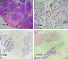
图7
施氏獭蛤雌雄同体组织切片 a—d. 雌性向雄性转换, 示雌雄生殖细胞共存同一滤泡(A)、雌雄滤泡并存同一性腺(B); c—d. 雌雄性腺共存, 精巢处于排放期, 卵巢处于休止期"

图7
| [1] |
蔡英亚, 劳赞, 陈东, 2005. 施氏獭蛤的生态观察[J]. 湛江海洋大学学报, 25(1): 39-42.
|
|
|
|
| [2] |
曹伏君, 刘永, 张春芳, 等, 2012. 施氏獭蛤 (Lutraria sieboldii) 性腺发育和生殖周期的研究[J]. 海洋与湖沼, 43(5): 976-982.
|
|
|
|
| [3] |
陈昭廷, 周洋, 顾志峰, 等, 2017. 海月水母(Aurelia aurita)精巢发育及精子的超微结构[J]. 海洋与湖沼, 48(1): 122-129.
|
|
|
|
| [4] |
丁汉波, 1987. 发育生物学[M]. 北京: 高等教育出版社: 55-60 (in Chinese).
|
| [5] |
顾海龙, 林志华, 沈伟良, 等, 2013. 泥蚶初级卵母细胞发育及卵黄发生的超微结构研究[J]. 海洋科学, 37(1): 49-53.
|
|
|
|
| [6] |
郭春阳, 徐善良, 2016. 蛤蜊科贝类的研究进展[J]. 生物学杂志, 33(1): 86-91.
|
|
|
|
| [7] |
韩龙江, 刘清华, 许飞, 等, 2017. 长牡蛎精子超低温冷冻后超微结构损伤研究[J]. 水生生物学报, 41(1): 220-227.
|
|
|
|
| [8] |
姜永华, 颜素芬, 2009. 中国龙虾卵子发生及卵黄发生的超微结构观察[J]. 中国水产科学, 16(5): 697-704.
|
|
|
|
| [9] |
姜永华, 颜素芬, 王重刚, 等, 2005. 凡纳滨对虾卵子发生的超微结构[J]. 水产学报, 29(4): 454-460.
|
|
|
|
| [10] |
焦宗垚, 刘永, 张春芳, 2010. 施氏獭蛤融合卵裂及其胚胎发育过程观察[J]. 动物学研究, 31(4): 408-414.
|
|
|
|
| [11] |
柯巧珍, 李琪, 闫红伟, 等, 2012. 山东北部沿海四角蛤蜊性腺发育年周期研究[J]. 中国海洋大学学报, 42(11): 28-34.
|
|
|
|
| [12] |
李霞, 2019. 水产动物组织胚胎学[M]. 北京: 中国农业出版社:178- 179.
|
|
|
|
| [13] |
栗志民, 刘志刚, 邓海东, 2011. 温度和盐度对企鹅珍珠贝清滤率、滤食率、吸收率的影响[J]. 水产学报, 35(1): 96-103.
|
|
|
|
| [14] |
连昌朋, 吴韬, 王超奇, 等, 2022. 广西北海营盘海域钝缀锦蛤(Tapes conspersus) 卵巢发育、卵子和卵黄发生的研究[J]. 热带海洋学报, 41(5): 170-179.
doi: 10.11978/2021185 |
|
|
|
| [15] |
刘超, 郭景兰, 彭张明, 等, 2015. 施氏獭蛤稚贝对高温和干露的耐受性研究[J]. 水产科学, 34(3): 169-173.
|
|
|
|
| [16] |
刘文广, 李琪, 高凤祥, 等, 2011. 长牡蛎繁殖周期、生化成分的季节变化与环境因子的关系[J]. 热带海洋学报, 30(3): 88-93.
|
|
|
|
| [17] |
刘志刚, 刘建勇, 刘付少梅, 2011. 不同潮位、密度及季节对皱肋文蛤中间培育效果的影响[J]. 海洋科学, 35(10): 34-41.
|
|
|
|
| [18] |
宁军号, 常亚青, 宋坚, 等, 2015. 偏顶蛤的性腺发育和生殖周期[J]. 中国水产科学, 22(3): 469-477.
|
|
|
|
| [19] |
潘彬斌, 李家乐, 白志毅, 2010. 池养三角帆蚌卵巢发育与卵子发生的组织学研究[J]. 上海海洋大学学报, 19(4): 452-456.
|
|
|
|
| [20] |
潘英, 秦小明, 潘红平, 2007. 大獭蛤软体部营养成分的分析与评价[J]. 广东海洋大学学报, 27(3): 78-81.
|
|
|
|
| [21] |
彭慧婧, 张守都, 郑德斌, 等, 2019. 施氏獭蛤全同胞家系建立及生长与存活性状分析[J]. 海洋科学, 43(7): 132-138.
|
|
|
|
| [22] |
阮飞腾, 高森, 李莉, 等, 2014. 山东沿海魁蚶繁殖周期与生化成分的周年变化[J]. 水产学报, 38(1): 47-55.
|
|
|
|
| [23] |
邵艳卿, 张炯明, 方军, 等, 2017. 人工蓄养斧文蛤的生殖周期及早期发育[J]. 中国水产科学, 24(1): 82-90.
|
|
doi: 10.3724/SP.J.1118.2017.16002 |
|
| [24] |
王斌, 栗志民, 刘志刚, 等, 2015. 施氏獭蛤室内规模化人工育苗技术研究[J]. 广东海洋大学学报, 35(1): 35-42.
|
|
|
|
| [25] |
王超奇, 徐炳杰, 吴韬, 等, 2023. 广西北部湾滩涂施氏獭蛤中培及养成期养殖密度比较研究[J]. 南方水产科学, 19(4): 105-115.
|
|
|
|
| [26] |
王梅芳, 余祥勇, 王君彦, 1999. 两种江珧雌雄同体及性转换现象[J]. 湛江海洋大学学报, 19(4): 6-10.
|
|
|
|
| [27] |
王如才, 王昭萍, 2008. 海水贝类养殖学[M]. 青岛: 中国海洋大学出版社:132- 133.
|
|
|
|
| [28] |
闻海波, 孙光兴, 丁图强, 等, 2020. 淮河橄榄蛏蚌繁殖类型与性腺发育观察[J]. 中国水产科学, 27(10): 1156-1166.
|
|
|
|
| [29] |
吴洪流, 2000. 波纹巴非蛤雌性生殖腺的组织学研究[J]. 海南大学学报, 18(3): 258-265.
|
|
|
|
| [30] |
吴洪流, 2002. 波纹巴非蛤性逆转时生殖腺的组织学变化[J]. 海洋科学, 26(1): 5-8.
|
|
|
|
| [31] |
吴明灿, 张立, 潘英, 等, 2015. 糙海参卵子发生及卵黄发生的超微结构[J]. 热带海洋学报, 34(3): 68-74.
doi: 10.11978/j.issn.1009-5470.2015.03.009 |
|
|
|
| [32] |
巫旗生, 文宇, 曾志南, 2017. 钝缀锦蛤繁殖周期和胚胎发育[J]. 中国水产科学, 24(3): 488-496.
|
|
doi: 10.3724/SP.J.1118.2017.16280 |
|
| [33] |
徐凤山, 张素萍, 2008. 中国海产双壳类图志[M]. 北京: 科学出版社:162-163.
|
|
|
|
| [34] |
徐红琴, 马慧妹, 曾起, 等, 2022. 温度对池蝶蚌雌雄同体和性逆转的影响[J]. 水生生物学报, 46(5): 741-753.
|
|
|
|
| [35] |
杨耀聪, 李复雪, 1994. 尖紫蛤生殖周期的研究[J]. 热带海洋, 13(2): 61-67.
|
|
|
|
| [36] |
应雪萍, 2002a. 泥螺卵子发生的超微结构研究[J]. 发育与生殖生物学报, 11(1): 29-36.
|
|
|
|
| [37] |
应雪萍, 2002b. 文蛤卵母细胞卵黄发生的超微结构[J]. 中国水产科学, 9(2): 125-128.
|
|
|
|
| [38] |
于非非, 余祥勇, 王梅芳, 等, 2007. 双壳类的性转换现象及其机理探讨[J]. 水生生物学报, 31(4): 576-580.
|
|
|
|
| [39] |
余祥勇, 王梅芳, 曹新云, 等, 2010. 斗嫁䗩卵子发生和雌性性腺组织学研究[J]. 水生生物学报, 34(5): 913-921.
|
|
|
|
| [40] |
种金豆, 李琪, 徐成勋, 等, 2020. 长牡蛎“海大3号”生长繁殖与营养成分的周年变化[J]. 水产学报, 44(3): 411-418.
|
|
|
|
| [41] |
周丽青, 杨爱国, 王清印, 等, 2014. 繁殖期雌雄同体虾夷扇贝生殖腺组织学观察[J]. 高技术通讯, 24(8): 874-880.
|
|
|
|
| [42] |
邹杰, 彭慧婧, 张守都, 等, 2020. 施氏獭蛤壳体表型性状对体质量的影响分析[J]. 水产科学, 39(4): 573-578.
|
|
|
|
| [43] |
doi: S0955-0674(17)30140-0 pmid: 29529563 |
| [44] |
doi: 10.1016/S0305-0491(99)00187-X |
| [45] |
doi: 10.1007/s11802-016-2855-6 |
| [46] |
doi: 10.1016/j.fishres.2013.10.003 |
| [47] |
doi: 10.1016/j.aquaculture.2006.10.028 |
| [48] |
|
| [49] |
doi: 10.1016/S1385-1101(03)00045-5 |
| [50] |
doi: 10.1016/0305-0491(73)90066-7 |
| [51] |
doi: 10.1016/j.aquaculture.2008.05.018 |
| [52] |
doi: 10.1016/j.aquaculture.2006.08.023 |
| [53] |
doi: 10.1016/S0990-7440(00)00135-2 |
| [54] |
doi: 10.1007/s11157-013-9310-6 |
| [55] |
doi: 10.1007/s12686-012-9774-7 |
| [56] |
doi: 10.1002/jmor.v281.8 |
| [1] | 徐炳杰, 刘一鸣, 邢清淦, 连昌朋, 吴韬, 潘英. 广西北部湾海域织锦巴非蛤精巢发育、精子发生及超微结构研究[J]. 热带海洋学报, 2024, 43(2): 59-68. |
| [2] | 徐炳杰, 刘一鸣, 连昌朋, 吴韬, 潘英. 广西北部湾海域织锦巴非蛤卵巢发育、卵子及卵黄发生的研究[J]. 热带海洋学报, 2024, 43(2): 48-58. |
| [3] | 吴韬, 潘英, 刘一鸣, 连昌朋, 徐炳杰, 王超奇, 杨凌. 广西北部湾海域施氏獭蛤精巢发育、精子发生及超微结构观察[J]. 热带海洋学报, 2024, 43(2): 69-80. |
| [4] | 连昌朋, 吴韬, 王超奇, 杨凌, 潘英. 广西北海营盘海域钝缀锦蛤(Tapes conspersus)卵巢发育、卵子和卵黄发生的研究[J]. 热带海洋学报, 2022, 41(5): 170-179. |
| [5] | 周欢, 林岗, 饶小珍. 刺巨藤壶精子的发生及其超微结构[J]. 热带海洋学报, 2020, 39(3): 98-105. |
| [6] | 宋悦凡, 曲翊, 曹旭鹏, 汪秋宽, 张卫. 南海小轴海绵的组织结构和细胞特征[J]. 热带海洋学报, 2016, 35(4): 71-81. |
| [7] | 吴明灿, 张立, 潘英, 黄光华, 李咏梅, 杨学明. 糙海参卵子发生及卵黄发生的超微结构[J]. 热带海洋学报, 2015, 34(3): 68-74. |
| [8] | 倪娜, 柳学周 徐永江, 赵明, 曲建忠. 雌性条斑星鲽脑垂体组织学观察[J]. 热带海洋学报, 2012, 31(6): 97-102. |
| [9] | 黄楷翔,吴瑞梹,陈弘成. 粗糙沼虾繁殖周期及其卵巢发育研究[J]. 热带海洋学报, 2011, 30(1): 159-164. |
| [10] | 黄建华,杨其彬,林黑着,陈旭,周发林,温为庚,江世贵. 南海野生斑节对虾亲虾卵巢发育过程中的脂肪酸组成[J]. 热带海洋学报, 2011, 30(1): 144-151. |
| [11] | 肖云朴,徐善良,孙敏,吕慧明. 黑鱾精子发生过程中的超微结构变化[J]. 热带海洋学报, 2011, 30(1): 107-112. |
| [12] | 曹伏君,罗杰,李长玲,刘楚吾. 细角螺的生殖系统组织学研究[J]. 热带海洋学报, 2010, 29(6): 57-64. |
| [13] | 许尤厚,刘学东,张吕平,胡超群. 凡纳滨对虾精子发生的超微结构研究[J]. 热带海洋学报, 2010, 29(4): 89-93. |
| [14] | 张殿彩,饶小珍,林岗,许友勤,陈寅山. 大竹蛏精子发生和精子的超微结构[J]. 热带海洋学报, 2009, 28(6): 131-135. |
|
||