
Journal of Tropical Oceanography >
Morphology and Phylogenetics study on the species of Amphidinium (Gymnodinials, Dinophyceae) from Weizhou Island, Guangxi
Received date: 2019-08-24
Request revised date: 2019-11-26
Online published: 2020-05-19
Supported by
National Nature Science Foundation of China(41576162)
National Nature Science Foundation of China(41876173)
Copyright
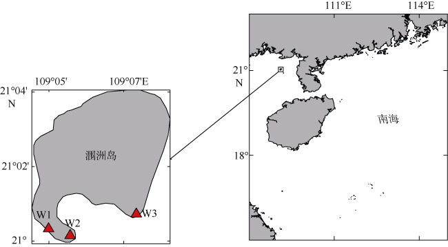
In this study, strains of Amphidinium from seaweeds and dead corals around Weizhou Island, Guangxi, China were isolated by single cell capillary pipette and cultured. The morphological features and phylogenetic analysis were determined based on light microscopy, scanning electron microscopy and molecular biology techiniques. Three Amphidinium species were identified, namely, Amphidinium operculatum, Amphidinium massertii and Amphidinium caeterae, among which A. operculatum and A. massertii were new records in China. The differences in morphology and phylogeny between different species were analyzed and compared. It was found that the morphological characteristics of the three Amphidinium species were significantly consistent with model species. This study adds information to the diversity and phylogenetic evolution of Amphidinium species in China, and extented the distribution of two new Amphidinium species to the coastal waters of southern China.
Key words: Weizhou Island; Amphidinium; morphology; phylogeny
JIANG Lichun , LI Qun , LÜ Songhui . Morphology and Phylogenetics study on the species of Amphidinium (Gymnodinials, Dinophyceae) from Weizhou Island, Guangxi[J]. Journal of Tropical Oceanography, 2020 , 39(3) : 106 -115 . DOI: 10.11978/2019077
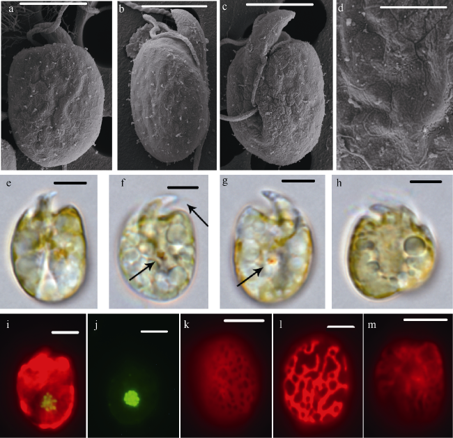
图2 具盖前沟藻扫描电镜图(a—d)与光镜图(e—l)a. 腹面观, 箭头指向横鞭毛孔; b. 背面观; c. 底面观; d. 二分裂细胞; e. 腹面观, 黑色箭头指向囊泡, 白色箭头指向眼点; f. 黑色箭头指向眼点; g. 细胞核; h. 叶绿体形状分布; i—k. 不同形状的具盖前沟藻; l. 二分裂细胞。各图比例尺为10μm Fig. 2 Scanning electron micrographs (a-d) and light micrographs (e-l) of A. operculatum. a) Ventral view; b) dorsal view; c) antapical view; d) binary division cell; e) ventral view, with black arrow pointing to the pusule and white arrow pointing to the eyespot; f) black arrow pointing to the eyespot; g) nucleus; h. distribution and shape of chloroplast; (i-k) A. operculatum with different shapes; l) binary division cell. Scale bars in a-l: 10 μm |
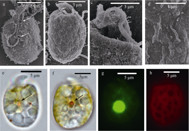
图3 玛氏前沟藻扫描电镜图(a—d)和光镜图(e—m)a. 背面观; b. 侧面观; c. 腹面观, 箭头指向横鞭毛孔; d. 细胞表面; e、f. 腹面观, 箭头指向蛋白核; g. 背面观, 箭头指向蛋白核; h. 二分裂细胞; i、j. 细胞核; k—m. 叶绿体形态分布; a—c、e—m比例尺为5μm, d为2μm Fig. 3 Scanning electron micrographs (a-d) and light micrographs (e-m) of A. massartii. a) Dorsal view; b) side view; c) ventral view, with arrow pointing to the pore of transverse flagellum; d) surface of cell; (e-f) ventral view, with arrow pointing to the pyrenoid; g) dorsal view, with arrow pointing to the pyrenoid; h) binary division cell; (i-j) nucleus; (k-m) distribution and shape of chloroplast. Scale bars in a-c and e-m: 5 μm; d: 2 μm |
表1 3个前沟藻种形态特征比较Tab. 1 Comparison of the morphological characters of three species of Amphidinium |
| 种名 | 长/μm | 宽/μm | 形状 | 纵鞭毛插入位置 | 蛋白核 | 泡 | 体鳞 |
|---|---|---|---|---|---|---|---|
| 具盖前沟藻(A.Operculatum) | 33~48 | 22~37 | 卵圆形 | 偏细胞底端1/3处 | 无 | 2个, 位于两个 鞭毛孔处 | 无 |
| 玛氏前沟藻(A.Massartii) | 10~16 | 7~12 | 卵圆形或椭圆形 | 细胞中部 | 1个, 细胞中部 | 无 | 有 |
| 强壮前沟藻(A.Carterae) | 12~19 | 9~14 | 卵圆形 | 细胞中部 | 1个, 细胞中部 | 无 | 无 |
图4 强壮前沟藻扫面电镜图(a—d)和光镜图(e—h)a. 腹面观, 箭头指向鞭毛孔; b. 背面观; c. 上锥部背面观; d. 细胞表面; e. 腹面观, 箭头指向蛋白核; f. 背面观, 箭头指向蛋白核; g. 细胞核; h. 叶绿体形态分布 Fig. 4 Scanning electron micrographs (a-d) and light micrographs (e-h) of A. carterae. a) Ventral view, with arrow pointing to the pore of flagellum; b) dorsal view; c) the dorsal view of epicone; d) surface of cell; e) ventral view, with arrow pointing to the pyrenoid; f) dorsal view, with arrow pointing to the pyrenoid; g) nucleus; h) the distribution and shape of chloroplast |
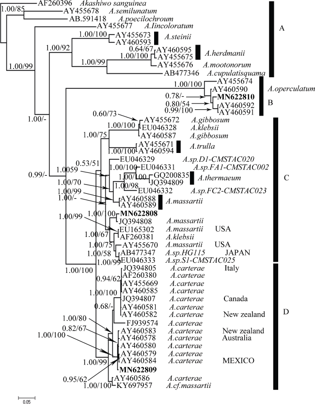
图5 前沟藻属LSU rDNA(D1-D3)的贝叶斯系统发育树节点数字代表贝叶斯分析法的后验概率(左)和最大似然法分析的置信值(右), 50%以下的置信值已省略 Fig. 5 Bayesian inference (BI) tree of the LSU rDNA (D1-D3 regions) of Amphidinium species. The number on the branch is the Posterior probabilities for BI analysis (left) and bootstrap values for maximum likelihood analysis (right). Support values below 50% have been omitted |
| 1 |
韩笑天, 颜天, 邹景忠 , 等, 2004. 强壮前沟藻(Amphidinium carterae Hulburt)形态特征及其生长特性研究[J]. 海洋与湖沼, 35(3):279-283.
|
| 2 |
张珍妮, 姜玥璐, 李曼璐 , 等, 2016. 强壮前沟藻(Amphidinium carterae Hulbert)在多重逆境条件下的生长和生理响应[J]. 海洋与湖沼, 47(5):982-989.
|
| 3 |
|
| 4 |
|
| 5 |
|
| 6 |
|
| 7 |
|
| 8 |
|
| 9 |
|
| 10 |
|
| 11 |
|
| 12 |
|
| 13 |
|
| 14 |
|
| 15 |
|
| 16 |
|
| 17 |
|
| 18 |
|
| 19 |
|
| 20 |
|
| 21 |
|
| 22 |
|
| 23 |
|
| 24 |
|
| 25 |
|
| 26 |
|
| 27 |
|
| 28 |
|
| 29 |
|
| 30 |
|
| 31 |
|
| 32 |
|
| 33 |
|
| 34 |
|
| 35 |
|
| 36 |
|
| 37 |
|
| 38 |
|
| 39 |
|
| 40 |
|
| 41 |
|
| 42 |
|
/
| 〈 |
|
〉 |

