Marine Biology

Antiviral chromones from the deep-sea hydrothermal-vent-derived Fusarium sp. SCSIO 06196

  • WANG Junfeng 1 ,
  • ZHOU Wenying 2 ,
  • TIAN Xinpeng 1 ,
  • LIU Yonghong , 1
Expand
  • 1. CAS Key Laboratory of Tropical Marine Bio-resources and Ecology, South China Sea Institute of Oceanology, Chinese Academy of Sciences, Guangzhou 510301, China
  • 2. Fifth School of Clinical Medicine, Guangzhou University of Chinese Medicine, Guangzhou 510405, China
LIU Yonghong. email:

Editor: YIN Bo

Received date: 2021-12-30

  Revised date: 2022-03-01

  Online published: 2022-03-21

Supported by

Science and Technology Project of Hainan Province(ZDKJ202018)

National Natural Science Foundation of China(21502204)

National Natural Science Foundation of China(41776169)

Abstract

Six chromone derivatives (1 ~ 6) were identified from the fungus Fusarium sp. SCSIO 06196 isolated from deep-sea hydrothermal vent environments in the Western Pacific. Their structures were elucidated mainly by extensive Nuclear Magnetic Resonance (NMR) spectroscopic, mass spectrometric (MS) and rotational analysis. Antiviral activities of the compounds 1 ~ 6 were tested. Among these compounds, compounds 1 ~ 3 exhibited significant in vitro anti-enterovirus 71 (EV-71) activities with IC50 values of 26.7, 19.8, and 22.0 μmol•L-1, respectively (IC50=177 μmol•L-1, Ribavirin). In addition, compound 1 showed weak antiviral activity against the H3N2 influenza virus with IC50 value of 8.6 μmol•L-1 (IC50=18.5 nmol•L-1, Tamiflu).

Cite this article

WANG Junfeng , ZHOU Wenying , TIAN Xinpeng , LIU Yonghong . Antiviral chromones from the deep-sea hydrothermal-vent-derived Fusarium sp. SCSIO 06196[J]. Journal of Tropical Oceanography, 2022 , 41(5) : 189 -193 . DOI: 10.11978/2021186

1 Introduction

Marine fungi from special ecological niches produce unique and biologically active secondary metabolites (Zhong et al, 2019; Ma et al, 2021). Within these vast ecological niches, one area remains particularly enigmatic: deep-sea hydrothermal ventscomprise one of the most unique environments on the planet. Owing to their extreme environmental conditions (high temperature, high pressure, sulfide- rich, low pH, and darkness), the microorganisms that live in or near these environments may be envisioned as containing diverse genetic and metabolic capabilities (Andrianasolo et al, 2009). As part of our investigations aimed at exploring bioactive secondary metabolites from fungal species inhabiting unique environments (Wang et al, 2016, 2015; Han et al, 2020), a fungal strain Fusarium sp. SCSIO 06196, isolated from deep-sea hydrothermal vent environments of the Western Pacific, was cultured in a nutrient-limited medium.

2 Results and discussion

Six chromone derivatives (1 ~ 6) were obtained and identified from fungal strain Fusarium sp. SCSIO 06196, isolated from deep-sea hydrothermal vent environments in the Western Pacific Ocean. The structures of the isolated compounds (1 ~ 6) were identified on the basis of spectral data and by comparing with those reported in the literature data. They were elucidated as epiremisporine B (1) (Xia et al, 2015), yicathin C (2) (Sun et al, 2013), citreorosein (3) (Fujimoto et al, 2004), physcion (4) (Zhang et al, 2003), janthinone (5) (Chai et al, 2012), pinselin (6) (Sun et al, 2013) (Figure 1).
Fig. 1 Structures of compounds 1 ~ 6 from the fungus Fusarium sp. SCSIO 06196
Cyclopentachromones (CPCs), the tricyclic cyclopentabenzopyan-9-one derivatives, are relatively rare in nature. Prior to this study, only a few in this family have been reported from fungi, including coniochaetones A-I, coniothyrione, diaportheones A-B, preussochromones D-F, cryptosporioptide, and remisporine A. Remisporine B is a unique CPC dimer that is resulted from a spontaneous Diels-Alder reaction of remisporine A (Kong et al, 2003; Xia et al, 2015). The 1H NMR spectrum of epiremisporine B (1) in DMSO-d6 showed two sets of 1H signals from the isomers 1a and 1b with ratio of 1 : 0.8. Significant differences in the 1H NMR data between 1a and 1b appeared mainly on the protons around C-2′ on the aliphatic rings, indicating that they are epimers at C-2′.
The antiviral (EV71 and H3N2) activities of compounds (1 ~ 6) were tested. Among these compounds, compounds 1 ~ 3 exhibited significant in vitro anti-enterovirus 71 activities with IC50 values of 26.7, 19.8, and 22.0 μmol·L-1, respectively (IC50 177 μmol·L-1 for Ribavirin). In addition, epiremisporine B (1) showed weak antiviral activity against the H3N2 influenza virus with IC50 value of 8.6 μmol·L-1 (IC50 18.5 nmol·L-1 for Tamiflu).

3 Experiment

3.1 General experimental procedures

1H, and 13C NMR, Distortionless Enhancement by Polarization Transfer (DEPT) spectra were recorded on the Bruker DRX-500 spectrometer (Bruker BioSpin, Fallanden, Switzerland) using TMS as internal standard and chemical shifts were recorded as δ-values. HRESIMS (including ESIMS) spectra were recorded on an Applied Biosystems Mariner 5140 spectrometer (Life Technologies Ltd, New York, USA). TLC and column chromatography were performed on plates precoated with silica gel GF254 (10 ~ 40 μm) and over silica gel (200 ~ 300 mesh) (Qingdao Marine Chemical Factory, Qingdao, China), and Sephadex LH-20 (Amersham Biosciences, Uppsala, Sweden), respectively. Analytical grade solvents were used in these experiments (Tianjin Fuyu Chemical and Industry Factory, Tianjin, China). Semipreparative HPLC was performed using an ODS column (YMC-pack ODS-A, 10 × 250 mm, 5 μm, 4 mL·min-1).

3.2 Fungal material

The fungus Fusarium sp. SCSIO 06196 was isolated from deep-sea hydrothermal vent environments collected in April 2014 from Western Pacific (126.8983°E, 27.7875°N) at a depth of 1028 m, and identified by the professor Xinpeng Tian, one of our co-authors. The fungus was identified using a molecular biological protocol by the sequencing of the ITS region. A BLAST search result showed that the sequence was mostly similar (100 %) to that of Fusarium sp. (compared to accession no. KT192277.1). A reference culture is deposited in our laboratory at -80 ℃. The fungus Fusarium sp. SCSIO 06196 was prepared on potato dextrose agar slants at 3.3 % salt concentration and stored at 4 ℃.

3.2.1 Culture conditions, extraction and isolation

Fusarium sp. SCSIO 06196 was cultured under static conditions at 26 ℃ for 60 days in one hundred 1000 mL conical flasks containing liquid medium (300 mL per flask) composed of starch soluble (10 g•L-1) and polypeptone (1 g•L-1), and tap water after adjusting its pH to 7.5. After 60 days, liquid medium from 100 flasks were collected and extracted using ethyl acetate (EtOAc). The filtrate was concentrated under vacuum to about a quarter of original volume and then extracted three times with EtOAc to give an EtOAc solution. The EtOAc solution was concentrated under vacuum to give an EtOAc extract (14.3 g). The EtOAc extract (14.3 g) was subjected to VLC on a silica gel column using step gradient elution with MeOH-CH2Cl2 (0 ~ 100 %) to separate into eight fractions based on TLC properties. Fraction 1 (0.9 g) was divided into six parts (Frs. 1-1 ~ 1-6) followed by HPLC (86 % MeOH / H2O). Fr.1-6 (56 mg) was further separated by HPLC (70 % MeOH / H2O) to yield 4 (6.2 mg, tR 19.7 min). Fraction 2 (2.1 g) was divided into five parts (Frs. 2-1 ~ 2-5) followed by Sephadex LH-20 (MeOH / CH2Cl2 = 1 : 1). Fr. 2-2 (0.4 g) was divided into six parts (Frs. 2-2-1 ~ 2-2-6) followed by HPLC (80 % MeOH / H2O). Fr. 2-2-5 (73 mg) was purified by HPLC (58 % CH3CN / H2O) to yield 1a / 1b (3.9 mg, tR 27.8 min). Fr. 2-3 (112 mg) was further purified by HPLC (70 % MeOH / H2O) to yield 6 (16.4 mg, tR 14.3 min), 2 (9.4 mg, tR 18.1 min) and 5 (7.2 mg, tR 21.9 min), respectively. Fraction 4 (1.6 g) was divided into three parts (Frs. 4-1 ~ 4-3) followed by Sephadex LH-20 (MeOH). Fr. 4-2 (106 mg) was further separated by HPLC (35 % CH3CN / H2O) to yield 3 (7.7 mg, tR 11.3 min).

3.2.2 Bioassay Protocols

EV71 was assayed on Vero cells with the CCK8 (DOjinDo, Japan) method. Vero cells (2 × 10³ cells•well-1) were seeded with DMEM medium (2 % FBS) into a 384-wellplate. After 24 h, 1000-fold serial dilution of the compound was added in triplicate to the 348-well plate. After incubation at 37 ℃ for 30 min, a two folds dilution 100 × the 50 % tissue culture infectious dose (TCID50) of EV71 virus in DMEM supplemented with 2 % FBS was added to each well. The plate was incubated at 37 ℃ for 72 ~ 96 h when the viral control cells showed complete CPE, the cell survival was quantified using CCK-8. The A450 of the well was measured with a microtiter plate reader (Envision, PerkinElmer) (Jia et al, 2019). The positive control was ribavirin with an IC50 value of 177 μmol•L-1.
The antiviral activity against H3N2 was evaluated by the CPE inhibition assay. Confluent MDCK cell monolayers were firstly incubated with influenza virus at 37 ℃ for 1 h. After removing the virus dilution, cells were maintained in infecting media (RPMI 1640, 4 μg•mL-1 of trypsin) containing different concentrations of test compounds. After 48 h at 37 ℃, the cells were fixed with 100 μL of 4 % formaldehyde for 20 min. After removal of the formaldehyde, the cells were stained with 0.1 % crystal violet for 30 min. The plates were washed and dried, and the intensity of crystal violet staining for each well was measured in a microplate reader (Bio-Rad, Hercules, CA, USA) at 570 nm. The IC50 value was calculated as the compound concentration required inhibiting influenza virus yield at 48 h post-infection by 50 %. Tamiflu was used as the positive control with IC50 value of 18.5 nmol•L-1, respectively.

3.3 The physicochemical data of the known compounds 1 ~ 6

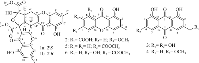
Epiremisporine B (1): a yellow amorphous powder (MeOH), $[\alpha ]_{D}^{25}$+ 419.5 (c 0.1 MeOH). ESI-MS m/z: 603 [M+Na]+; CD (MeOH) Δε (nm): +13.01 (206.0), +1.95 (221), 0 (227), -1.94 (238), 0 (242), +12.57 (260), 0 (271), -6.36 (282), 0 (298), +4.24 (332), 0 (439). (S)-epiremisporine B (1a): 1H NMR (DMSO-d6, 500 MHz): δ: 3.78 (1H, t, J = 9.0 Hz, H-3), 4.99 (1H, d, J = 9.0 Hz, H-4), 6.83 (1H, s, H-8), 6.66 (1H, s, H-10), 2.31 (3H, s, H-15), 3.75 (3H, s, H-16), 3.13 (1H, m, H-3°), 2.70 (1H, m, H-4°a), 2.60 (1H, m, H-4°b), 6.94 (1H, s, H-8°), 6.73 (1H, s, H-10°), 2.39 (3H, s, H-15°), 3.74 (3H, s, H-16°), 12.16 (1H, s, 11-OH), 7.86 (1H, s, 2°-OH), 12.51 (1H, s, 11°-OH); 13C NMR (DMSO-d6, 125 MHz): δ: 170.8 (C, C-1), 88.2 (C, C-2), 47.0 (CH, C-3), 36.9 (CH, C-4), 169.3 (C, C-5), 156.8 (C, C-7), 108.4 (CH, C-8), 147.5 (C, C-9), 112.4 (CH, C-10), 159.6 (C, C-11), 108.4 (C, C-12), 178.9 (C, C-13), 119.2 (C, C-14), 21.5 (CH3, C-15), 52.8 (CH3, C-16), 169.5 (C, C-1°), 105.7 (C, C-2°), 42.7 (CH, C-3°), 26.3 (CH, C-4°), 168.1 (C, C-5°), 155.6 (C, C-7°), 107.7 (CH, C-8°), 147.5 (C, C-9°), 112.4 (CH, C-10°), 159.8 (C, C-11°), 107.7 (C, C-12°), 179.5 (C, C-13°), 111.9 (C, C-14°), 21.8 (CH3, C-15°), 52.4 (CH3, C-16°). (R)-epiremisporine B (1b): 1H NMR (DMSO-d6, 500 MHz): δ: 3.89 (1H, t, J = 8.5 Hz, H-3), 5.02 (1H, d, J = 9.0 Hz, H-4), 6.81 (1H, s, H-8), 6.67 (1H, s, H-10), 2.30 (3H, s, H-15), 3.74 (3H, s, H-16), 2.79 (1H, m, H-3°), 2.48 (1H, m, H-4°a), 2.43 (1H, m, H-4°b), 6.91 (1H, s, H-8°), 6.73 (1H, s, H-10°), 2.39 (3H, s, H-15°), 3.75 (3H, s, H-16°), 12.13 (1H, s, 11-OH), 7.53 (1H, s, 2°-OH), 12.49 (1H, s, 11°-OH); 13C NMR (DMSO-d6, 125 MHz): δ: 171.4 (C, C-1), 89.4 (C, C-2), 46.6 (CH, C-3), 36.2 (CH, C-4), 168.2 (C, C-5), 156.8 (C, C-7), 108.4 (CH, C-8), 147.5 (C, C-9), 112.4 (CH, C-10), 159.9 (C, C-11), 108.4 (C, C-12), 178.9 (C, C-13), 118.7 (C, C-14), 21.5 (CH3, C-15), 52.6 (CH3, C-16), 167.7 (C, C-1°), 106.2 (C, C-2°), 47.3 (CH, C-3°), 27.0 (CH, C-4°), 167.5 (C, C-5°), 155.6 (C, C-7°), 108.4 (CH, C-8°), 147.5 (C, C-9°), 112.0 (CH, C-10°), 159.6 (C, C-11°), 107.7 (C, C-12°), 179.4 (C, C-13°), 111.7 (C, C-14°), 21.8 (CH3, C-15°), 52.3 (CH3, C-16°).
Yicathin C (2): ESI-MS m/z: 301 [M+H]+; 1H NMR (DMSO-d6, 500 MHz): δ: 6.61 (1H, s, H-2), 6.83 (1H, s, H-4), 6.85 (1H, s, H-5), 6.79 (1H, s, H-7), 2.37 (3H, s, H-11), 3.86 (3H, s, H-13), 12.73 (1H, s, 8-OH); 13C NMR (DMSO-d6, 125 MHz): δ: 160.5 (C, C-1), 111.1 (CH, C-2), 148.5 (CH, C-3), 107.3 (CH, C-4), 155.2 (C, C-4a), 157.8 (C, C-10a), 103.2 (CH, C-5), 134.9 (C, C-6), 113.4 (CH, C-7), 168.5 (C, C-8), 108.4 (C, C-8a), 178.9 (C, C-9), 105.7 (C, C-9a), 22 (CH3, C-11), 52.7 (CH3, C-13).
Citreorosein (3): ESI-MS m/z: 287 [M+H]+; 1H NMR (DMSO-d6, 500 MHz): δ: 7.36 (1H, s, H-2), 7.76 (1H, s, H-4), 7.21 (1H, s, H-5), 6.64 (1H, s, H-7), 4.73 (2H, s, H-11); 13C NMR (DMSO-d6, 125 MHz): δ: 161.4 (C, C-1), 120.8 (CH, C-2), 152.7 (CH, C-3), 117.0 (CH, C-4), 133.0 (C, C-4a), 108.3 (CH, C-5), 164.7 (C, C-6), 108.0 (CH, C-7), 167.1 (C, C-8), 109.8 (C, C-8a), 189.1 (C, C-9), 114.2 (C, C-9a), 181.6 (C, C-10), 135.1 (C, C-10a), 62.1 (CH3, C-11).
Physcion (4): ESI-MS m/z: 307 [M+Na]+; 1H NMR (CDCl3, 500 MHz): δ: 6.7 (1H, s, H-7), 3.96 (3H, s, H-15), 2.47 (3H, s, H-16), 12.3 (1H, s, 1-OH), 8.65 (1H, s, 8-OH); 13C NMR (CDCl3, 125 MHz): δ: 162.6 (C, C-1), 124.7 (CH, C-2), 148.6 (CH, C-3), 121.4 (CH, C-4), 108.4 (CH, C-5), 166.7 (C, C-6), 107.9 (CH, C-7), 165.3 (C, C-8), 190.9 (C, C-9), 182.2 (C, C-10), 133.3 (C, C-11), 113.8 (CH3, C-12), 110.4 (C, C-13), 135.4 (C, C-14), 22.3 (CH3, C-15), 56.2 (CH3, C-16).
Janthinone (5): ESI-MS m/z: 285 [M+H]+; 1H NMR (DMSO-d6, 500 MHz): δ: 7.47 (1H, d, J = 7.3 Hz, H-2), 7.83 (1H, t, J = 8.0 Hz, H-3), 7.57 (1H, d, J = 8.5 Hz, H-4), 6.17 (1H, s, H-5), 6.64 (1H, s, H-7), 3.99 (3H, s, H-11), 2.27 (3H, s, H-12), 8.65 (1H, s, 8-OH); 13C NMR (DMSO-d6, 125 MHz): δ: 133.5 (C, C-1), 122.5 (CH, C-2), 134.9 (CH, C-3), 119.2 (CH, C-4), 155.4 (C, C-4a), 155.2 (C, C-4b), 107.2 (CH, C-5), 149.3 (C, C-6), 111.2 (CH, C-7), 160.8 (C, C-8), 106.4 (C, C-8a), 180.1 (C, C-9), 116.7 (C, C-9a), 168.8 (C, C-10), 52.3 (CH3, C-11), 21.7 (CH3, C-12).
Pinselin (6): ESI-MS m/z: 301 [M+H]+; 1H NMR (DMSO-d6, 500 MHz): δ: 7.47 (1H, s, H-3), 7.59 (1H, d, J = 9.0 Hz, H-4), 6.86 (1H, s, H-5), 6.63 (1H, s, H-7), 6.64 (1H, s, H-7), 2.38 (3H, s, H-11), 3.85 (3H, s, H-12), 12.18 (1H, s, 8-OH); 13C NMR (DMSO-d6, 125 MHz): δ: 120.1 (C, C-1), 150.9 (C, C-2), 125.4 (CH, C-3), 117.0 (CH, C-4), 149.3 (C, C-4a), 155.4 (C, C-4b), 107.4 (CH, C-5), 149.3 (C, C-6), 106.0 (CH, C-7), 160.4 (C, C-8), 110.8 (C, C-8a), 180.3 (C, C-9), 117.2 (C, C-9a), 166.9 (C, C-10), 22.1 (CH3, C-11), 52.3 (CH3, C-12).

4 Conclusion

To discover the bioactive natural compounds produced by the fungus from deep-sea hydrothermal vent environments, six compounds (1 ~ 6), were discovered from Fusarium sp. SCSIO 06196, isolated from deep-sea hydrothermal vent environments of the Western Pacific. Among them, compounds 1 ~ 3 exhibited significant in vitro anti-enterovirus 71 activities with IC50 values of 26.7, 19.8, and 22.0 μmol•L-1, respectively (IC50 177 μmol•L-1 for the positive control Ribavirin).
[1]
ANDRIANASOLO E H, HARAMATY L, ROSARIO- PASSAPERA R, et al, 2009. Ammonificins A and B, hydroxyethylamine chroman derivatives from a cultured marine hydrothermal vent bacterium, Thermovibrio ammonificans[J]. Journal of Natural Products, 72(6): 1216-1219.

DOI

[2]
CHAI YUNJING, CUI CHENGBIN, LI CHANGWEI, et al, 2012. Activation of the dormant secondary metabolite production by introducing gentamicin-resistance in a marine-derived Penicillium purpurogenum G59[J]. Marine Drugs, 10(12): 559-582.

DOI

[3]
FUJIMOTO H, NAKAMURA E, OKUYAMA E, et al, 2004. Six immunosuppressive features from an ascomycete, Zopfiella longicaudata, found in a screening study monitored by immunomodulatory activity[J]. Chemical & Pharmaceutical Bulletin, 52(8): 1005-1008.

[4]
HAN WENRONG, CAI JIA, ZHONG WEIMAO, et al, 2020. Protein tyrosine phosphatase 1B (PTP1B) inhibitors from the deep-sea fungus Penicillium chrysogenum SCSIO 07007[J]. Bioorganic Chemistry, 96: 103646.

DOI

[5]
JIA QIAN, DU YUQI, WANG CHEN, et al, 2019. Azaphilones from the marine sponge-derived fungus Penicillium sclerotiorum OUCMDZ-3839[J]. Marine Drugs, 17(5): 260.

DOI

[6]
KONG FANGMING, CARTER G T, 2003. Remisporine B, a novel dimeric chromenone derived from spontaneous Diels-Alder reaction of remisporine A[J]. Tetrahedron Letters, 44(15): 3119-3122.

DOI

[7]
MA LILI, TIAN XINPENG, LI GUIJU, et al, 2021. Research status and development trends of natural products from marine microorganisms[J]. Journal of Tropical Oceanography, 40(5): 134-146. (in Chinese with English abstract)

DOI

[8]
SUN KUNLAI, WANG YI, FU PENG, et al, 2013a. Studies on the secondary metabolites of Eurotium herbariorum HT-2 symbiotic with Enteromorpha prolifera[J]. Chinese Journal of Marine Drugs, 32(1): 37-45. (in Chinese with English abstract)

[9]
SUN RANRAN, MIAO FENGPING, ZHANG JING, et al, 2013b. Three new xanthone derivatives from an algicolous isolate of Aspergillus wentii[J]. Magnetic Resonance in Chemistry, 51(1): 65-68.

DOI

[10]
WANG JUNFENG, WEI XIAOYI, QIN XIAOCHU, et al, 2015. Arthpyrones A-C, pyridone alkaloids from a sponge-derived fungus Arthrinium arundinis ZSDS1-F3[J]. Organic Letters, 17(3): 656-659.

DOI

[11]
WANG JUNFENG, WEI XIAOYI, QIN XIAOCHU, et al, 2016. Antiviral merosesquiterpenoids produced by the Antarctic fungus Aspergillus ochraceopetaliformis SCSIO 05702[J]. Journal of Natural Products, 79(1): 59-65.

DOI

[12]
XIA MINGWEN, CUI CHENGBIN, LI CHANGWEI, et al, 2015. Rare chromones from a fungal mutant of the marine-derived Penicillium purpurogenum G59[J]. Marine Drugs, 13(8): 5219-5236.

DOI

[13]
ZHANG WEN, LOU HONGXIANG, LI GUANGYAO, et al, 2003. A new triterpenoid from Entodon okamurae Broth[J]. Journal of Asian Natural Products Research, 5(3): 189-195.

DOI

[14]
ZHONG WEIMAO, WANG JUNFENG, WEI XIAOYI, et al, 2019. Three pairs of new spirocyclic alkaloid enantiomers from the marine-derived fungus Eurotium sp. SCSIO F452[J]. Frontiers in Chemistry, 7: 350.

DOI

Outlines

/