Exploitation of Marine Resources

Intracellular and extracellular metabolites analysis and key metabolite screening on the Bacillus paranthracis SG49

  • LIU Shuai ,
  • LIU Xuerui ,
  • ZHANG Rui ,
  • GUO Xiangrui ,
  • YU Zhen ,
  • SUN Hao ,
  • ZHANG Yanying
Expand
  • Ocean School, Yantai University, Yantai 264005, China
ZHANG Yanying. email:

Copy editor: SUN Cuici

Received date: 2024-03-23

  Revised date: 2024-04-03

  Online published: 2024-04-28

Supported by

NSFC-Shandong Joint Fund(U2106208)

National Natural Science Foundation of China(41976147)

Abstract

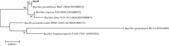
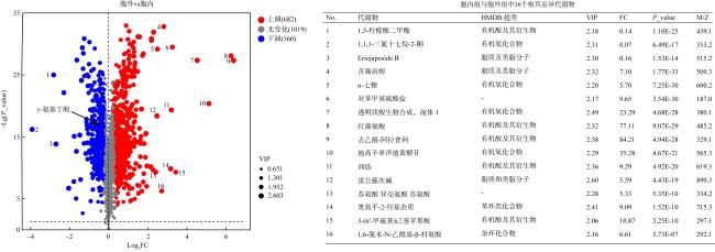
As a result of climate change and human intervention, jellyfish outbreaks have become a serious ecological disaster that threatens coastal economies and marine ecosystems. Globally, there is an urgent need to prevent jellyfish blooms. Microorganisms play an important role in the growth and development of marine invertebrates. Co-culture experiments revealed that Bacillus paranthracis SG49 inhibits the settlement and metamorphosis of Aurelia coerulea planula larvae. The key metabolic pathways and mechanisms behind this inhibition, however, require further investigation. Using non-targeted metabolomics technology, the intracellular and extracellular metabolites of SG49 were detected, differences between the two groups were analyzed, and potential metabolites affecting planula larval metamorphosis were identified. Our results showed that SG49 intracellular and extracellular metabolites were significantly different. Specifically, seven substances were screened for their potential inhibitory activities, including 3-hydroxy-2-oxindole, kanamycin, apramycin, streptomycin, streptomycin sulfate, gallic acid, and coniferyl alcohol. Bacterial biofilms and microorganism growth can be inhibited by these metabolites. Our findings provide a theoretical basis and strain resources to prevent jellyfish outbreaks in the future.

Cite this article

LIU Shuai , LIU Xuerui , ZHANG Rui , GUO Xiangrui , YU Zhen , SUN Hao , ZHANG Yanying . Intracellular and extracellular metabolites analysis and key metabolite screening on the Bacillus paranthracis SG49[J]. Journal of Tropical Oceanography, 2025 , 44(1) : 122 -132 . DOI: 10.11978/2024068

近年来, 由于海洋表面温度的上升、富营养化和过度捕捞等因素, 近岸海域灾害水母频繁暴发(Dong, 2019; Rowe et al, 2022)。海月水母(Aurelia coerulea)广泛分布于南纬40°至北纬70°的沿海水域, 是海洋中最丰富的水母群之一(Uye et al, 2005; Purcell et al, 2007; Yoon et al, 2019)。在过去的几年中, 海月水母在黄海北部、胶州湾和台湾海峡的暴发现象更加频繁(Wang et al, 2015, 2023b), 暴发后会堵塞沿海工业冷却系统, 破坏渔网捕捞, 对发电厂和渔业产生不利影响, 阻碍沿岸经济发展(Dong et al, 2010; Delannoy et al, 2011; Báez et al, 2022)。
海洋无脊椎动物在其生命周期中通常经历附着变态阶段。浮浪幼虫一旦完成浮游阶段, 就会定居在合适的基质上。随后, 幼虫发生形态变化并变形为与成体相似的幼体(Hadfield, 2011; Mayorova et al, 2012; Brekhman et al, 2015; Hoyer et al, 2024)。附着和变态阶段对海洋无脊椎动物的生存和分布至关重要(He et al, 2021)。其中涉及许多复杂过程(Chandramouli et al, 2014; Hadfield et al, 2021; Rischer et al, 2022), 受多重因素影响, 包括物理因素(如温度、盐度、光、溶解氧、金属阳离子和附着底物)、化学因素(如分泌物、神经递质、不饱和脂肪酸)和生物因素(如生物本身的生理状态)(Roberts et al, 2007; Feng et al, 2010; Alfaro et al, 2014; Franco et al, 2016; Cavalcanti et al, 2020)。抑制灾害水母浮浪幼虫附着变态发育, 可以缩小水螅体定植蔓延的范围, 降低灾害水母种群的数量。近年来, 水母暴发已成为近岸生态灾害问题, 从防生物污损角度出发, 利用生物手段抑制灾害水母浮浪幼虫附着变态, 预防水母灾害暴发, 对海洋生态系统健康发展具有重要意义(Pinteus et al, 2020; Thomé et al, 2022)。
海洋微生物资源丰富, 并能够产生多种代谢产物, 同时具备可以对来源生物和代谢产物进行遗传和化学修饰的可能性等特性, 使其成为活性次级代谢产物的重要来源。近年来, 从海洋微生物中发现了很多次生代谢产物, 包括从微生物中提取的萜类、类固醇、类胡萝卜素、酚类、呋喃酮、生物碱、多肽和内酯等化合物均具有抑制海洋无脊椎动物幼虫附着变态的活性(Satheesh et al, 2016)。如链霉菌(Streptomyces albidoflavus) UST040711-291和链霉菌(Streptomyces sp.) GWS-BW-H5产生的丁烯内酯, 能有效抑制纵条纹藤壶(Balanus amphitrite)幼虫的附着(Dickschat et al, 2005; Xu et al, 2010)。链霉菌UST040711-290产生的12-甲基十四烷酸(12-MTA), 能够抑制多毛类秀丽隐杆线虫的幼虫附着(Li et al, 2006; Dobretsov et al, 2013)。此外, 微生物还能通过分泌蛋白酶抑制海洋无脊椎动物幼虫附着变态发育, 如依氏假交替单胞菌(Pseudoalteromonas issachenkonii)产生的蛋白酶能够抑制苔藓虫幼虫的附着(Dobretsov et al, 2007)。芽孢杆菌是常见的可以产生次级代谢产物和蛋白酶的细菌(Belousova et al, 2021), 可以产生胰蛋白酶、枯草杆菌蛋白酶等中性和碱性的蛋白酶类和胞外多糖(König, 2006), 其中, 蛋白酶和胞外多糖已被证明有抑制海洋无脊椎动物附着变态(Dobretsov et al, 2007; Peng et al, 2020; Liang et al, 2021; Li et al, 2022; Hu et al, 2024), 这些研究结果为深入研究微生物抑制海洋无脊椎物幼虫附着变态的机制提供了重要参考。
我们在前期研究中发现菌株SG49与海月水母浮浪幼虫共培养时, 海月水母浮浪幼虫不能发生附着变态现象, 推测其通过分泌胞外代谢产物抑制海月水母浮浪幼虫的附着变态, 本研究利用代谢组学技术, 分析菌株胞内外代谢物的差异, 筛选关键抑制性代谢产物, 并分析其生物学功能。研究结果为抑制水母幼虫的附着变态提供了新的见解, 为利用微生物资源抑制灾害水母浮浪幼虫附着变态, 解决灾害水母暴发现象提供理论依据。

1 材料与方法

1.1 菌株、培养基和试剂

菌株的分离鉴定: 在山东威海天鹅湖海域采集日本鳗草根系沉积物, 称量2g沉积物于10ml无菌离心管中, 加入8mL无菌海水, 置于28℃摇床中180 r·min-1培养24h。4℃、6000g离心15min收集上清液, 吸取200µL上清液于多种液体培养基中置于28℃摇床中180r·min-1培养, 再取200µL菌液于相应的固体培养基中涂布(菌株SG49是在2216E固体培养基中分离鉴定), 进行多次纯化划线后提取DNA寄送生工生物工程(上海)股份有限公司鉴定, 获得16S rDNA序列。
菌株活化: 将保存于-80℃超低温冰箱中的菌株SG49接种于2216E固体培养基上, 放置28℃恒温培养箱中培养两天。用接种针挑取单菌落接种至200mL的2216E液体培养基中, 置于28℃摇床中180r·min-1培养。待菌液长至对数生长期, 4℃、6000g离心15min收集菌体, 使用无菌生理盐水漂洗后, 制成菌悬液。
2216E培养基: 胰蛋白胨5g, 酵母提取物1g, 二水磷酸铁0.1g, 氯化钠25g, 去离子水1000mL, pH为7.6~7.8, 固体培养基再添加15g琼脂。胰蛋白胨、酵母提取物、二水磷酸铁、氯化钠、氢氧化钠、琼脂等试剂均为工业用化学纯产品。

1.2 菌体和上清液样品采集

细菌菌体的收集: 将菌株SG49接种到2216E液体培养基中180r·min-1培养3d, 每4h测量菌液的光密度值, 绘制生长曲线, 在对数生长期离心收集菌体。将100mL的菌液分两次转移至50mL无菌旋盖尖底离心管中, 室温下6000g离心15min, 加入1.5mL无菌生理盐水重悬。将菌液转移至1.5mL无菌离心管中, 4℃下14000g离心5min收集菌体, 将~3g菌体在干冰条件下寄送美吉生物医药科技有限公司(中国, 上海)进行代谢物检测。
上清液的获取: 将对数生长期的400mL菌液分别转移至8个50mL无菌旋盖尖底离心管中, 4℃下6000g离心15min, 收集上清。将离心管开口用无菌锡纸包裹后用无菌牙签扎6~8个孔, 立于冷冻干燥机中冻干72~84h至干燥粉末状, 将冻干样品寄送至美吉生物(中国, 上海)进行代谢物检测。

1.3 代谢物检测

参考Sun等的方法, 从菌株SG49菌体和上清液粉末中提取和检测代谢物(Sun et al, 2023)。将约3g菌体和上清液粉末与400µL甲醇:水溶液按4:1(体积比)的比例混合。混合液在-20℃下孵育, 并用高通量组织破碎机Wonbio-96c (上海万柏生物技术有限公司) 处理6min, 5℃涡旋30s, 40kHz超声处理30min。混合液在-20℃孵育30min沉淀蛋白质, 13000g离心15min。收集上清液后进行液相色谱-质谱(LC-MS)分析。从8个菌体样本和8个上清液样本中取等量混合制备成3个混合质控 (quality control, QC) 样品。QC样品与菌体和上清液粉末按照相同的程序进行处理和测试, 以监测稳定性。采用Thermo UHPLC-Q Exactive HF-X系统进行样品的LC-MS/MS检测, 配备ACQUITY HSS T3色谱柱(100m×2.1mm, 1.8µm; Waters, USA), 样品从HSS T3柱中分离3µL, 进行质谱检测, 检测过程在上海美吉生物完成。

1.4 代谢物分析

运用软件Progenesis QI (Waters Corporation, Milford, USA) 对液相色谱-质谱(LC-MS)的初始数据集进行处理, 具体包括基线过滤、峰识别、积分处理、保留时间的调整以及峰对齐等。得到一个综合了保留时间、质荷比(M/Z)和峰值强度信息的数据矩阵。将质谱数据(MS)及串联质谱数据(MS/MS)与公共代谢物数据库HMDB(The Human Metabolome Database, https://hmdb.ca/)和Metlin进行匹配分析, 以识别和确认代谢物信息。

1.5 信号通路分析及关键性差异代谢产物的筛选

对数据矩阵进行预处理, 保留在80%以上的样品中检测到的代谢物, 对低于检测限的样品设定其值为最低代谢物值填补空缺值。代谢物采用总和归一化法进行处理, 获得归一化后的数据矩阵。以QC数据作为内标, 剔除相对标准偏差 (relative standard deviation, RSD)>30%的数据, 并进行log10对数转换。
为了深入探究不同样本之间代谢物组成的差异, 采用R语言中的“ropls”包(版本1.6.2)进行正交最小偏二乘判别分析(orthogonal partial least squares-discriminant analysis, OPLS-DA)。通过200次循环交叉验证, 对模型的稳健性进行严格评估, 检验OPLS-DA模型的拟合程度。在此基础上, 根据VIP(variable important for the projection)值(VIP>1)以及student’s t检验的P值(P<0.05)来筛选具有统计学意义的差异代谢物。
利用KEGG数据库对差异代谢物进行注释, 获得生物学功能。利用Python的“scipy.stats”包进行通路富集分析, 利用Fisher精确检验确定其最相关的生物学途径。

2 结果

2.1 菌株SG49的16S rDNA进化树

对菌株SG49做16S系统进化树分析(图1), 发现其近缘序列为Bacillus paranthracis Mn5T (MACE01000012), 相似度为99.51%。该菌株隶属于厚壁菌门(Firmicutes), 芽孢杆菌纲(Bacilli), 芽孢杆菌目(Bacillales), 芽孢杆菌科(Bacillaceae), 芽孢杆菌属(Bacillus), 副炭疽芽孢杆菌(Bacillus paranthracis)。
图1 菌株 SG49 的系统进化发育树

Fig. 1 The phylogenetic tree of strain SG49

2.2 代谢组数据的质控

通过对低质量峰进行过滤、填充、筛选和数据转换后, 共获得了14453个代谢物, 其中2061个代谢物在数据库中得到注释, 包括阳离子代谢物1134个, 阴离子代谢物895个(表1)。经处理代谢物质量提高, 在RSD<0.3条件下其代谢物峰所占比值从95.4%升至98.2%, 而且处理前后峰所占的累积比例>70%, 表明该代谢物数据能够较好的反映实际情况, 数据质量合格(图2a)。样本的代谢物聚类分析结果表明, 胞内和胞外代谢产物具有显著差别, 平行样本之间具有较好的一致性, 表明结果可靠, 可信度高(图2b)。
表1 菌株SG49胞内和胞外代谢物丰度

Tab. 1 The abundance of intracellular and extracellular metabolites from SG49

离子模式 所有峰值 已鉴定代谢物 Library数据库代谢物 KEGG数据库代谢物
阳离子 5618 1134 1004 497
阴离子 8835 927 895 454
图2 代谢物数据质量分析

a. QC样本评估曲线, 虚线表示预处理前数据, 实线表示预处理后数据; b. 样本相关性热图

Fig. 2 The data quality analysis of all metabolites. (a) The quality evaluation curve of QC samples. Dashed lines indicate pre-pretreatment data and solid lines represent post-pretreatment data. (b) The heat map of sample correlations

2.3 代谢组样本比较分析

利用OPLS-DA对菌株SG15胞内和胞外的代谢产物的构成模式进行分析, 结果表明, SG49菌体和上清液中的阴离子和阳离子代谢物能够明显区分开, 组内代谢物的组成模式较为统一(图3a、b)。第一预测主成分(component 1)解释度占阳离子代谢物变化的91.30%(图3a), 阴离子代谢物变化的91.00%(图3b)。第一正交成分(orthogonal component 1)解释度占阳离子代谢变化的0.89%(图3a), 阴离子代谢变化的1.22%(图3b)。通过200次置换检验对OPLS-DA模型进行验证, 发现回归曲线的截距为<0, 表明模型没有过拟合(图3c、d)。此外, 在阳离子模式下R2XR2YQ2分别为0.913、1.000和1.000, 阴离子模式下R2XR2YQ2分别为0.910、1.000和0.999, 这也证实了上述OPLS-DA模型的可靠性。
图3 菌株SG49胞内外代谢物OPLS-DA及置换检验图

a. 阳离子模式下胞内组和胞外组的OPLA-DA分析; b. 阴离子模式下胞内组和胞外组的OPLA-DA分析; c. 阳离子模式下OPLA-DA分析的置换检验; d. 阴离子模式下OPLA-DA分析的置换检验

Fig. 3 The OPLS-DA plot of metabolites in SG49 intracellular and extracellular groups. (a) OPLA-DA analysis of intracellular and extracellular groups in positive ion mode; (b) OPLA-DA analysis of intracellular and extracellular groups in negative ion mode; (c) permutation testing for OPLA-DA analysis in positive ion mode; (d) permutation testing for OPLA-DA analysis in negative ion mode

2.4 差异代谢物及胞外代谢物注释信息

我们对SG49菌体及上清中共有的及特有的代谢物进行比较分析(图4e), 发现胞内组和胞外组有1570个共有代谢物, 胞内组有123个特有代谢物, 胞外组有382个特有代谢物。我们对胞内组和胞外组特有的代谢产物进行分析, HMDB化合物分类结果表明, 胞内组在HMDB数据库中鉴定出108个代谢物, 占胞内总注释代谢物的87.80%(图4a)。其中有机酸及其衍生物(31.48%)是最丰富的代谢物, 其次是脂质和类脂分子(28.70%)、有机杂环化合物(13.89%)和有机氧化合物(7.41%)。胞外组在HMDB数据库中鉴定出362个代谢物, 占胞外总注释代谢物的94.76%(图4c)。其中有机酸及其衍生物(33.43%)是最丰富的代谢物, 其次是脂质和类脂分子(17.96%)、有机杂环化合物(14.92%)和有机氧化合物(10.22%)。在组成比例上细菌菌体和上清液中的代谢物类别较为相似, 但上清液中特有的代谢产物数目比菌体特有的高。我们利用KEGG通路富集分析研究不同组特有代谢产物的富集情况, 发现胞内组中氨基酸代谢通路显著富集, 包括甘氨酸、丝氨酸和苏氨酸代谢、半胱氨酸和蛋氨酸代谢、丙氨酸、天冬氨酸和谷氨酸代谢和精氨酸生物合成等通路(图4b)。在胞外组中, KEGG通路富集结果显示氨基酸代谢通路同样活跃, 包括β-丙氨酸代谢、精氨酸和脯氨酸代谢、氰基氨基酸代谢和缬氨酸、亮氨酸和异亮氨酸生物合成等代谢通路(图4d)。除此之外, 胞外组的富集通路中还存在极为重要的多种次生代谢产物的生物合成通路。
图4 代谢物注释及富集分析

a. 胞内超类水平上HMDB注释的特有代谢物; b. 胞内特有代谢物KEGG通路富集分析图; c. 胞外超类水平上HMDB注释的特有代谢物; d. 胞外特有代谢物KEGG通路富集分析图; e. 胞外和胞内的样本比较Venn分析图

Fig. 4 The annotation and enrichment analysis of metabolites. (a) Unique metabolites of intracellular group at superclass level of HMDB database; (b) enrichment analysis diagram of KEGG pathway of unique metabolites in intracellular group; (c) unique metabolites of extracellular group at superclass level of HMDB database; (d) enrichment analysis diagram of KEGG pathway of unique metabolites in extracellular groups; (e) Venn analysis chart comparing samples from intracellular and extracellular groups
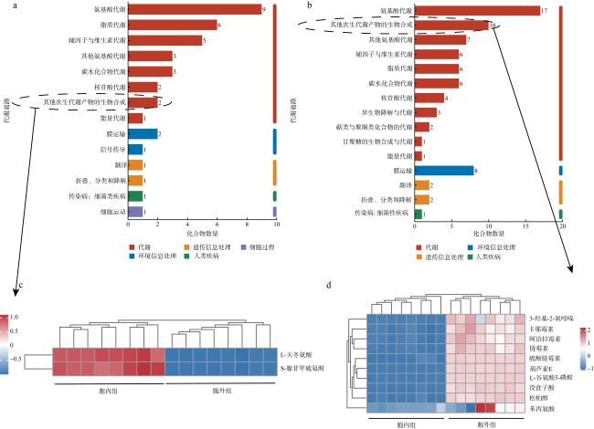
2.5 关键差异代谢产物分析

为了分析SG49菌体和上清液之间的差异代谢物, 我们筛选了基于VIP>1(OPLS-DA), P<0.05 (student’s t检验)和差异表达倍数(fold change, FC)>1或FC<1的代谢物。共获得2015种阳离子代谢物和3300种阴离子代谢物, 其中360种代谢物上调, 682种代谢物下调 (图5)。在HMDB和Metlin数据库中注释到了530种阳离子代谢物和512种阴离子代谢物。为了进一步分析SG49菌体和上清液之间的差异代谢物, 我们基于VIP>2, P<0.05和FC>5或FC<5进行了代谢物筛选, 共获得37种阳离子代谢物和99种阴离子代谢物, 其中13种代谢物上调, 3种代谢物下调。其中5种阳离子代谢物和11种阴离子代谢物在HMDB和Metlin数据库中注释到。
图5 菌株SG49胞内外差异代谢物分析的火山图(左图)和16个胞内与胞外组显著差异代谢物(右图)

Fig. 5 Volcanic map of differential metabolite analysis of SG49 intracellular and extracellular groups (left panel) and 16 significantly different metabolites between intracellular and extracellular groups (right panel)

2.6 胞内外代谢物KEGG功能注释

为研究菌株SG49影响海月水母附着变态过程的关键代谢产物, 我们对胞内外特有的代谢物分别进行KEGG功能通路注释(图6)。胞内组胞内代谢物共获得14条KEGG功能通路(图6a), 其中, 氨基酸代谢通路中的代谢物丰度最高, 其他依次是脂质代谢、辅助因子和维生素的代谢和碳水化合物代谢。胞外组胞外代谢物共获得15条KEGG功能通路(图6b), 其中, 氨基酸代谢通路的代谢物丰度最高, 其次是其他次生代谢物的生物合成、辅助因子和维生素的代谢、脂质代谢和碳水化合物代谢等。将两组中与次级代谢产物相关的其他次生代谢物的生物合成组分单独分析研究其在胞内外丰度变化情况, 我们发现胞内组只有2种物质(图6c), 分别是S-腺苷甲硫氨酸和L-天冬氨酸。胞外组有10种物质(图6d), 分别是3-羟基-2-氧吲哚、卡那霉素、安普霉素、链霉素、硫酸链霉素、松柏醇、苯丙氨酸、葫芦素E、没食子酸和L-谷氨酸5-磷酸, 除苯丙氨酸外, 其他9种物质与胞内组丰度差异明显。
图6 胞内外特有代谢物的KEGG功能通路注释

a. 胞内组特有代谢物的KEGG功能通路; b. 胞外组特有代谢物的KEGG功能通路; c. 胞内组中其他次生代谢物的生物合成组物质的丰度变化; d. 胞外组中其他次生代谢物的生物合成组物质的丰度变化

Fig. 6 KEGG functional pathway of intracellular and extracellular unique metabolites. (a) KEGG pathway of unique metabolites in intracellular group; (b) KEGG pathway of unique metabolites in extracellular group; (c) the abundance of other secondary metabolite biosynthesis in intracellular group; (d) the abundance of other secondary metabolite biosynthesis in extracellular group

3 讨论

在海洋无脊椎动物早期发育过程中通常会经历从浮游状态到底栖生物状态的附着过程和变态发育, 包括幼虫的内部结构、外部形态、生理过程和行为的发育(Shen et al, 2018)。本课题组利用微生物与水母幼虫共培养实验, 获得能够抑制海月水母浮浪幼虫附着变态的菌株SG49, 经分类鉴定, 该菌株隶属于厚壁菌门(Firmicutes), 芽孢杆菌纲(Bacilli), 芽孢杆菌目 (Bacillales), 芽孢杆菌科(Bacillaceae), 芽孢杆菌属(Bacillus), 副炭疽芽孢杆菌种(Bacillus paranthracis)。芽孢杆菌属在海洋无脊椎动物幼虫的附着和变态过程发挥重要作用, 如芽孢杆菌属中的海水芽孢杆菌(Bacillus aquimaris)具有与海洋无脊椎动物幼虫附着变态相关的收缩结构, 其形成的生物被膜可以诱导秀丽隐杆线虫(Hydroides elegans)幼虫的附着过程(Freckelton et al, 2017)。深海环境中的芽孢杆菌生物被膜可以产生胞外多糖, 能够诱导厚壳贻贝(Mytilus coruscus)幼虫的附着和变态(Chang et al, 2021)。菌株SG49与海月水母浮浪幼虫共培养时, 水母浮浪幼虫不会发生附着变态现象, 揭示菌株SG49能够显著抑制海月水母浮浪幼虫的附着变态。已有研究揭示副炭疽芽孢杆菌种细胞呈革兰氏染色阳性, 兼性厌氧, 不运动, 杆状, 在2017年被Liu等人归类为蜡样芽孢杆菌类群(Bacillus cereus) (Liu et al, 2017)。
副炭疽芽孢杆菌具有产酶(刘佳慧 等, 2023; Tshisikhawe et al, 2023)、抗菌(Diale et al, 2021)、生物降解(Du et al, 2023)等功能, 在工业生产上有巨大潜力。从海绵表面分离到的蜡样芽孢杆菌, 能够抑制生物被膜细菌和微藻的粘附(Satheesh et al, 2012), Jin等人研究了海绵细菌的粗提物对硅藻的抗粘附活性, 揭示芽孢杆菌可能是抗污染化合物的潜在来源(Jin et al, 2014)。然而, 副炭疽芽孢杆菌抑制海洋无脊椎动物附着变态相关的代谢产物研究较少, 其抑制海洋无脊椎动物附着变态的机制仍然未知。
菌株SG49胞内和胞外代谢产物存在显著差异, 差异代谢产物主要包括有机酸及其衍生物、脂质与类脂分子和有机杂环化合物。在有机酸及其衍生物类别中, γ-氨基丁酸(GABA)是一种神经递质, 可诱导浮游红鲍幼虫(Haliotis rufescens)和东泥织纹螺幼虫(Ilyanassa obsoleta)附着并发生变态(Morse et al, 1979; Biscocho et al, 2018), γ-氨基丁酸在菌株SG49胞内具有丰富的表达量, 而胞外含量很少, 推测菌株SG49可能是通过调控γ-氨基丁酸的释放来抑制海月水母附着变态过程。游离脂肪酸如棕榈烯酸、亚油酸或花生四烯酸, 可作为诱导剂参与幼虫变态(Hu et al, 2021)。在海藻表面的假单胞菌通过产生有机酸异丙嗪-1-羧酸, 显著抑制海洋细菌、藻类孢子和藤壶幼虫附着变态(Burgess et al, 2003)。链霉菌属UST040711-290菌株产生12-甲基十四烷酸(12-MTA), 能够显著抑制多毛类秀丽隐杆线虫的幼虫附着行为(Xu et al, 2009)。因此, 微生物产生的有机酸及其衍生物与生物的附着和变态行为密切相关。菌株SG49胞内与胞外特有代谢物中, 有机酸及其衍生物含量较高, 推测有机酸及其衍生物可能在抑制海月水母浮浪幼虫附着变态的过程中发挥作用。
菌株胞内外的差异代谢产物主要富集在氨基酸代谢和次级代谢产物的生物合成。在本研究中, 菌株的色氨酸代谢、缬氨酸、亮氨酸和异亮氨酸生物合成、精氨酸和脯氨酸代谢和组氨酸代谢等氨基酸代谢途径显著富集。氨基酸代谢不仅支撑蛋白质合成, 还涉及能量产生、核苷酸形成及氧化还原平衡, 维持细胞和生物体功能(黄文明 等, 2024), 在海洋无脊椎动物的生存和发育中起着重要的作用。目前, 微生物通过产生氨基酸抑制海洋无脊椎动物幼虫附着变态相关的研究较少。
已有研究揭示, 水母的浮浪幼虫在无菌环境下无法正常附着, 微生物合成的寡肽是诱导浮浪幼虫的附着变态发育的关键物质(Hofmann et al, 1978; Kroiher et al, 1999)。此外, 无菌环境下海月水母的螅状幼体也无法进行横裂生殖(Peng et al, 2023)。海月水母的基因组结果也证实其不能合成色氨酸, 依赖共生微生物提供(Gold et al, 2019)。因此, 水母幼虫的变态发育与微生物息息相关。在菌株SG49胞外代谢产物中, 显著富集的次生代谢物包括3-羟基-2-氧吲哚、卡那霉素、安普霉素、链霉素、硫酸链霉素、松柏醇、苯丙氨酸、葫芦素E、没食子酸和L-谷氨酸5-磷酸。根据之前的研究表明, 这些代谢物大多与抑制细菌的生物被膜形成或抑制微生物生长有关(Almeida et al, 2017; Khan et al, 2020; Mansouri et al, 2021; Atlas et al, 2023; Wang et al, 2023a)。推测副炭疽芽孢杆菌SG49通过产生次级代谢产物抑制水母浮浪幼虫附着变态相关的微生物, 从而抑制海月水母浮浪幼虫的附着和变态过程。生物被膜是一种附着在生物体表面并被细胞外基质或细胞外聚合物(EPS)包裹的结构化微生物群落(Weiland-Bräuer et al, 2020)。这些微生物组分形成稳定的结构和理化梯度, 有利于共生微生物与宿主之间的基因转移, 加强细胞之间的信息交流(Cavalcanti et al, 2020)。生物被膜中的细菌分泌的溶血磷脂、胞外多糖或其他水溶性小分子物质能够诱导幼虫启动变态发育(Guo et al, 2017, 2021; Li et al, 2021)。利用抗生素来控制生物被膜的形成能够抑制幼虫附着变态(Atlas et al, 2023)。安普霉素对多种细菌种类具有抗菌活性, 可以有效抑制铜绿假单胞菌和肺炎克雷伯菌生物被膜形成能力(Atlas et al, 2023)。卡那霉素可与司托霉素发生协同作用, 显著抑制单核增生乳杆菌的生物被膜形成(Wang et al, 2023a)。链霉素能够显著抑制铜绿假单胞菌生物被膜形成活性, 降低铜绿假单胞菌群体感应(quorum sensing)相关毒力因子的产生, 并抑制铜绿假单胞菌的溶血活性(Khan et al, 2020)。硫酸链霉素对金黄色葡萄球菌、大肠杆菌和铜绿假单胞菌的生物被膜形成均有抑制作用(Mansouri et al, 2021)。没食子酸是一种多酚类化合物, 具有高抗菌活性(Abdelsalam et al, 2022)。没食子酸过硫酸盐能够有效抑制地中海贻贝(Mytilus galloprovincialis)的附着(Almeida et al, 2017; Parisi et al, 2022)。因此, 菌株SG49可能通过产生这些抑制海月水母浮浪幼虫共生微生物的生物被膜形成的代谢产物来抑制幼虫的附着和变态过程。但是这些代谢产物的作用机理还不清楚, 有待进一步深入研究。

4 结论

水母暴发已成为近岸生态灾害问题, 利用生物手段防治水母灾害对沿海城市社会经济稳定发展具有重要意义。芽孢杆菌SG49通过释放代谢产物抑制海月水母浮浪幼虫的附着变态, 经过对菌株SG49代谢组学的研究, 发现菌株SG49胞内外代谢物主要包括有机酸及其衍生物、有机杂环化合物与脂质和类脂分子, 菌株SG49代谢产物的生物学功能主要集中在氨基酸代谢和次级代谢产物的生物合成。研究筛选出7个具有潜在抑制活性的物质, 分别为3-羟基-2-氧吲哚、卡那霉素、安普霉素、链霉素、硫酸链霉素、没食子酸和松柏醇, 这些代谢产物可能通过抑制水母共附生细菌生物被膜形成或抑制微生物生长, 达到抑制水母浮浪幼虫附着变态的效果, 但具体的抑制机制需要进一步研究。
[1]
黄文明, 尹梦丽, 陈煜, 等, 2024. 一株高效同化氨氮霉菌的筛选及其代谢组分析[J]. 微生物学报, 64(1): 161-173.

HUANG WENMING, YIN MENGLI, CHEN YU, et al, 2024. Screening and metabolomic analysis of a fungal strain efficiently assimilating ammonia nitrogen[J]. Acta Microbiologica Sinica, 64(1): 161-173 (in Chinese with English abstract).

[2]
刘佳慧, 吕红, 林娟, 2023. 一株产低温碱性淀粉酶蕈状芽孢杆菌的分离筛选和发酵优化[J]. 四川大学学报(自然科学版), 60(2): 026001.

LIU JIAHUI, HONG, LIN JUAN, 2023. Isolation and screening of psychrophilic and alkaline amylase-producing Bacillus mycoides strain 3F1 and the optimum fermentation for enzyme production[J]. Journal of Sichuan University (Natural Science Edition), 60(2): 026001 (in Chinese with English abstract).

[3]
ABDELSALAM K M, SHALTOUT N A, IBRAHIM H A, et al, 2022. A comparative study of biosynthesized marine natural-product nanoparticles as antifouling biocides[J]. Oceanologia, 64(1): 35-49.

[4]
ALFARO A C, YOUNG T, BOWDEN K, 2014. Neurophysiological control of swimming behaviour, attachment and metamorphosis in black-footed abalone (Haliotis iris) larvae[J]. New Zealand Journal of Marine and Freshwater Research, 48(3): 314-334.

[5]
ALMEIDA J R, CORREIA-DA-SILVA M, SOUSA E, et al, 2017. Antifouling potential of nature-inspired sulfated compounds[J]. Scientific Reports, 7: 42424.

[6]
ATLAS N, UZAIR B, MOVELLAN J, et al, 2023. In vitro activity of novel apramycin-dextran nanoparticles and free apramycin against selected Dutch and Pakistani Klebsiella pneumonia isolates[J]. Heliyon, 9(12): e22821.

[7]
BÁEZ J C, PENNINO M G, ALBO-PUIGSERVER M, et al, 2022. Effects of environmental conditions and jellyfish blooms on small pelagic fish and fisheries from the Western Mediterranean Sea[J]. Estuarine, Coastal and Shelf Science, 264: 107699.

[8]
BELOUSOVA M E, MALOVICHKO Y V, SHIKOV A E, et al, 2021. Dissecting the environmental consequences of Bacillus thuringiensis application for natural ecosystems[J]. Toxins, 13(5): 355.

[9]
BISCOCHO D, COOK J G, LONG J, et al, 2018. GABA is an inhibitory neurotransmitter in the neural circuit regulating metamorphosis in a marine snail[J]. Developmental Neurobiology, 78(7): 736-753.

[10]
BREKHMAN V, MALIK A, HAAS B, et al, 2015. Transcriptome profiling of the dynamic life cycle of the scypohozoan jellyfish Aurelia aurita[J]. BMC Genomics, 16: 74.

[11]
BURGESS J G, BOYD K G, ARMSTRONG E, et al, 2003. The development of a marine natural product-based antifouling paint[J]. Biofouling, 19(S1): 197-205.

[12]
CAVALCANTI G S, ALKER A T, DELHERBE N, et al, 2020. The influence of bacteria on animal metamorphosis[J]. Annual Review of Microbiology, 74: 137-158.

[13]
CHANDRAMOULI K H, QIAN PEIYYUAN, RAVASI T, 2014. Proteomics insights: proteins related to larval attachment and metamorphosis of marine invertebrates[J]. Frontiers in Marine Science, 1: 52.

[14]
CHANG RUIHENG, YANG LITING, LUO MING, et al, 2021. Deep-sea bacteria trigger settlement and metamorphosis of the mussel Mytilus coruscus larvae[J]. Scientific Reports, 11(1): 919.

[15]
DELANNOY C M J, HOUGHTON J D R, FLEMING N E C, et al, 2011. Mauve stingers (Pelagia noctiluca) as carriers of the bacterial fish pathogen Tenacibaculum maritimum[J]. Aquaculture, 311(1-4): 255-257.

[16]
DIALE M O, KAYITESI E, SEREPA-DLAMINI M H, 2021. Genome in silico and in vitro analysis of the probiotic properties of a bacterial endophyte, Bacillus paranthracis strain MHSD3[J]. Frontiers in Genetics, 12: 672149.

[17]
DICKSCHAT J S, MARTENS T, BRINKHOFF T, et al, 2005. Volatiles released by a Streptomyces species isolated from the North Sea[J]. Chemistry & Biodiversity, 2(7): 837-865.

[18]
DOBRETSOV S, ABED R M M, TEPLITSKI M, 2013. Mini-review: Inhibition of biofouling by marine microorganisms[J]. Biofouling, 29(4): 423-441.

[19]
DOBRETSOV S, XIONG HAIRONG, XU YING, et al, 2007. Novel antifoulants: inhibition of larval attachment by proteases[J]. Marine Biotechnology, 9(3): 388-397.

[20]
DONG ZHIJUN, 2019. Blooms of the moon jellyfish Aurelia: causes, consequences and controls[M]// SHEPPARD C. World seas: an environmental evaluation. 2nd ed. New York: Academic Press: 163-171.

[21]
DONG ZHIJUN, LIU DONGYAN, KEESING J K, 2010. Jellyfish blooms in China: Dominant species, causes and consequences[J]. Marine Pollution Bulletin, 60(7): 954-963.

[22]
DU XINYUE, JIANG YAO, SUN YAWEN, et al, 2023. Biodegradation of Inosine and Guanosine by Bacillus paranthracis YD01[J]. International Journal of Molecular Sciences, 24(19): 14462.

[23]
FENG DANQING, KE CAIHUAN, LU CHANGYI, et al, 2010. The influence of temperature and light on larval pre-settlement metamorphosis: a study of the effects of environmental factors on pre-settlement metamorphosis of the solitary ascidian Styela canopus[J]. Marine and Freshwater Behaviour and Physiology, 43(1): 11-24.

[24]
FRANCO S C, ALDRED N, CRUZ T, et al, 2016. Modulation of gregarious settlement of the stalked barnacle, Pollicipes pollicipes: a laboratory study[J]. Scientia Marina, 80(2): 217-228.

[25]
FRECKELTON M L, NEDVED B T, HADFIELD M G, 2017. Induction of invertebrate larval settlement; different bacteria, different mechanisms?[J]. Scientific Reports, 7: 42557.

[26]
GOLD D A, KATSUKI T, LI YANG, et al, 2019. The genome of the jellyfish Aurelia and the evolution of animal complexity[J]. Nature Ecology & Evolution, 3(1): 96-104.

[27]
GUO HUIJUAN, RISCHER M, SPERFELD M, et al, 2017. Natural products and morphogenic activity of γ-Proteobacteria associated with the marine hydroid polyp Hydractinia echinata[J]. Bioorganic and Medicinal Chemistry, 25(22): 6088-6097.

[28]
GUO HUIJUAN, RISCHER M, WESTERMANN M, et al, 2021. Two distinct bacterial biofilm components trigger metamorphosis in the colonial hydrozoan Hydractinia echinata[J]. mBio, 12(3): e0040121.

[29]
HADFIELD M G, 2011. Biofilms and marine invertebrate larvae: What bacteria produce that larvae use to choose settlement sites[J]. Annual Review of Marine Science, 3: 453-470.

[30]
HADFIELD M G, FRECKELTON M L, NEDVED B T, 2021. The natural sequence of events in larval settlement and metamorphosis of Hydroides elegans (Polychaeta; Serpulidae)[J]. PLoS One, 16(5): e0249692.

[31]
HE JIAN, WU ZHIWEN, CHEN LIYING, et al, 2021. Adenosine triggers larval settlement and metamorphosis in the mussel Mytilopsis sallei through the ADK-AMPK-FoxO pathway[J]. ACS Chemical Biology, 16(8): 1390-1400.

[32]
HOFMANN D K, NEUMANN R, HENNE K, 1978. Strobilation, budding and initiation of scyphistoma morphogenesis in the rhizostome Cassiopea andromeda (Cnidaria: Scyphozoa)[J]. Marine Biology, 47(2): 161-176.

[33]
HOYER J, KOLAR K, ATHIRA A, et al, 2024. Polymodal sensory perception drives settlement and metamorphosis of Ciona larvae[J]. Current Biology, 34(6): 1168-1182.e7.

[34]
HU XIAOMENG, WANG XIAOYU, PENG LIHUA, et al, 2024. Decline of induction capability by outer membrane vesicles on larval metamorphosis of mussels through enzymatic treatments[J]. International Biodeterioration and Biodegradation, 190: 105783.

[35]
HU XIAOMENG, ZHANG JUNBO, DING WENYANG, et al, 2021. Reduction of mussel metamorphosis by inactivation of the bacterial thioesterase gene via alteration of the fatty acid composition[J]. Biofouling, 37(8): 911-921.

[36]
JIN CUILI, XIN XIAYING, YU SIYU, et al, 2014. Antidiatom activity of marine bacteria associated with sponges from San Juan Island, Washington[J]. World Journal of Microbiology & Biotechnology, 30(4): 1325-1334.

[37]
KHAN F, LEE J W, PHAM D T N, et al, 2020. Streptomycin mediated biofilm inhibition and suppression of virulence properties in Pseudomonas aeruginosa PAO1[J]. Applied Microbiology and Biotechnology, 104(2): 799-816.

[38]
KÖNIG H, 2006. Bacillus species in the intestine of termites and other soil invertebrates[J]. Journal of Applied Microbiology, 101(3): 620-627.

[39]
KROIHER M, BERKING S, 1999. On natural metamorphosis inducers of the cnidarians Hydractinia echinata (Hydrozoa) and Aurelia aurita (Scyphozoa)[J]. Helgoland Marine Research, 53(2): 118-121.

[40]
LI JIAZHENG, ZHANG CHI, HU XIAOMENG, et al, 2022. Impact of different enzymes on biofilm formation and mussel settlement[J]. Scientific Reports, 12(1): 4685.

[41]
LI MINGYU, WANG KAI, JIA CHENZHENG, et al, 2021. Bacteroidetes bacteria, important players in the marine sponge larval development process[J]. iScience, 24(6): 102662.

[42]
LI XIANCUI, DOBRETSOV S, XU YING, et al, 2006. Antifouling diketopiperazines produced by a deep-sea bacterium, Streptomyces fungicidicus[J]. Biofouling, 22(3): 187-194.

[43]
LIANG XIAO, ZHANG JUNBO, SHAO ANQI, et al, 2021. Bacterial cellulose synthesis gene regulates cellular c-di-GMP that control biofilm formation and mussel larval settlement[J]. International Biodeterioration and Biodegradation, 165: 105330.

[44]
LIU YANG, DU JUAN, LAI QILIANG, et al, 2017. Proposal of nine novel species of the Bacillus cereus group[J]. International Journal of Systematic and Evolutionary Microbiology, 67(8): 2499-2508.

[45]
MANSOURI M, KHAYAM N, JAMSHIDIFAR E, et al, 2021. Streptomycin sulfate-loaded niosomes enables increased antimicrobial and anti-biofilm activities[J]. Frontiers in Bioengineering and Biotechnology, 9: 745099.

[46]
MAYOROVA T D, KOSEVICH I A, MELEKHOVA O P, 2012. On some features of embryonic development and metamorphosis of Aurelia aurita (Cnidaria, Scyphozoa)[J]. Russian Journal of Developmental Biology, 43(5): 271-285.

[47]
MORSE D E, HOOKER N, DUNCAN H, et al, 1979. γ-aminobutyric acid, a neurotransmitter, induces planktonic abalone larvae to settle and begin metamorphosis[J]. Science, 204(4391): 407-410.

[48]
PARISI C, SANDONNINI J, COPPOLA M R, et al, 2022. Biocide vs. eco-friendly antifoulants: role of the antioxidative defence and settlement in Mytilus galloprovincialis[J]. Journal of Marine Science and Engineering, 10(6): 792.

[49]
PENG LIHUA, LIANG XIAO, CHANG RUIHENG, et al, 2020. A bacterial polysaccharide biosynthesis-related gene inversely regulates larval settlement and metamorphosis of Mytilus coruscus[J]. Biofouling, 36(7): 753-765.

[50]
PENG SAIJUN, YE LIJING, LI YONGXUE, et al, 2023. Microbiota regulates life-cycle transition and nematocyte dynamics in jellyfish[J]. iScience, 26(12): 108444.

[51]
PINTEUS S, LEMOS M F L, FREITAS R, et al, 2020. Medusa polyps adherence inhibition: A novel experimental model for antifouling assays[J]. Science of the Total Environment, 715: 136796.

[52]
PURCELL J E, UYE S I, LO W T, 2007. Anthropogenic causes of jellyfish blooms and their direct consequences for humans: a review[J]. Marine Ecology Progress Series, 350: 153-174.

[53]
RISCHER M, GUO HUIJUAN, BEEMELMANNS C, 2022. Signalling molecules inducing metamorphosis in marine organisms[J]. Natural Product Reports, 39(9): 1833-1855.

[54]
ROBERTS R D, KAWAMURA T, HANDLEY C M, 2007. Factors affecting settlement of abalone (Haliotis iris) larvae on benthic diatom films[J]. Journal of Shellfish Research, 26(2): 323-334.

[55]
ROWE C E, KEABLE S J, AHYONG S T, et al, 2022. Physiological responses of the upside-down jellyfish, Cassiopea (Cnidaria: Scyphozoa: Cassiopeidae) to temperature and implications for their range expansion along the east coast of Australia[J]. Journal of Experimental Marine Biology and Ecology, 554: 151765.

[56]
SATHEESH S, BA-AKDAH M A, AL-SOFYANI A A, 2016. Natural antifouling compound production by microbes associated with marine macroorganisms—A review[J]. Electronic Journal of Biotechnology, 21: 26-35.

[57]
SATHEESH S, SONIAMBY A R, SHANKAR C V S, et al, 2012. Antifouling activities of marine bacteria associated with sponge (Sigmadocia sp.)[J]. Journal of Ocean University of China, 11(3): 354-360.

[58]
SHEN MINGHUI, DI GUILAN, LI MIN, et al, 2018. Proteomics studies on the three larval stages of development and metamorphosis of Babylonia areolata[J]. Scientific Reports, 8(1): 6269.

[59]
SUN HAO, XIE ZIQIANG, YANG XIAOZHOU, et al, 2023. New insights into microbial and metabolite signatures of coral bleaching[J]. Science of the Total Environment, 892: 164258.

[60]
THOMÉ P E, RIVERA-ORTEGA J, 2022. Specific antibacterial activity against potential pathogens and restraining of larvae settlement from a pigmented Pseudoalteromonas strain isolated from the jellyfish Cassiopea xamachana[J]. International Aquatic Research, 14(2): 95-105.

[61]
TSHISIKHAWE M L, DIALE M O, ABRAHAMS A M, et al, 2023. Screening and production of industrially relevant enzymes by Bacillus paranthracis strain MHDS3, a potential probiotic[J]. Fermentation, 9(11): 938.

[62]
UYE S, SHIMAUCHI H, 2005. Population biomass, feeding, respiration and growth rates, and carbon budget of the scyphomedusa Aurelia aurita in the Inland Sea of Japan[J]. Journal of Plankton Research, 27(3): 237-248.

[63]
WANG WEI, LIU JUNSHENG, ZHOU JINWEI, et al, 2023a. Synergistic effect of kanamycin and amikacin with setomimycin on biofilm formation inhibition of Listeria monocytogenes[J]. Microbial Pathogenesis, 185: 106447.

[64]
WANG XIAOCHENG, JIN QINGQING, YANG LU, et al, 2023b. Aggregation process of two disaster-causing jellyfish species, Nemopilema nomurai and Aurelia coerulea, at the intake area of a nuclear power cooling-water system in Eastern Liaodong Bay, China[J]. Frontiers in Marine Science, 9: 1098232.

[65]
WANG YANTAO, SUN SONG, 2015. Population dynamics of Aurelia sp. 1 ephyrae and medusae in Jiaozhou Bay, China[J]. Hydrobiologia, 754(1): 147-155.

[66]
WEILAND-BRÄUER N, PINNOW N, LANGFELDT D, et al, 2020. The native microbiome is crucial for offspring generation and fitness of Aurelia aurita[J]. mBio, 11(6): e02336-20.

[67]
XU YING, HE HONGPING, SCHULZ S, et al, 2010. Potent antifouling compounds produced by marine Streptomyces[J]. Bioresource Technology, 101(4): 1331-1336.

[68]
XU YING, LI HONGLEI, LI XIANCUI, et al, 2009. Inhibitory effects of a branched-chain fatty acid on larval settlement of the polychaete Hydroides elegans[J]. Marine Biotechnology, 11(4): 495-504.

[69]
YOON E A, LEE K, CHAE J, et al, 2019. Density estimates of moon jellyfish (Aurelia coerulea) in the Yeongsan estuary using nets and hydroacoustics[J]. Ocean Science Journal, 54(3): 457-465.

Outlines

/