Marine Biology

Development of testis and ultrastructure of spermatozoa in Mastigias papua

  • ZHANG Ruixue , 1, 2 ,
  • YANG Wenhao 1, 2 ,
  • CHEN Nan , 1, 2 ,
  • WANG Shuhong 1, 2, 3
Expand
  • 1. Fisheries College of Jimei University, Xiamen 361021, China
  • 2. Ornamental Aquarium Engineering Research Centre in University of Fujian Province, Xiamen 361021, China
  • 3. Key Laboratory of Healthy Mariculture for the East China Sea, Ministry of Agriculture and Rural Affairs, Xiamen 361021, China
CHEN Nan. email:

Editor: LIN Qiang

Received date: 2024-01-02

  Revised date: 2024-02-08

  Online published: 2024-02-28

Supported by

Natural Science Foundation of Xiamen, China(3502Z202372021)

Research Start-up Funds of Jimei University(ZQ2021023)

Abstract

In this study, Mastigias Papua was selected as the sample, its testis development and spermatozoa ultrastructure was observed in great detail. The development phases of testis and the ultrastructure of the spermatozoa were also studied. The results showed that the nest of M. papus with a bell diameter of (4.99±0.07)cm gradually became matured after 26d-40d under experimental conditions, and the spermatozoa bundle could be continuously discharged for 2-3 months. According to the structural characteristics of spermatozoa follicles and spermatogenesis, the development of the testis can be divided into three stages, namely stage I: the outer part of the testis is not visible to naked eye, the seminal vesicles are small and transparent, and are arranged loosely, and there is no cavity inside; Phase II: the testis is circular, milky white, with gradient development of the gonads. The seminal vesicles that are far from the gastric filament are larger and more tightly arranged, with an internal cavity; Stage III: The folds of the testis are clearly sac-shaped, and under light microscopy the seminal vesicles at different developmental stages can be seen. A large number of seminal vesicles are filled with mature spermatozoa, present in spermbundles. The spermatozoa of M. papus was (50.64±3.93)μm in length, including a sharp chilli shape head (7.61±0.63)μm, a slightly protruding middle section (1.00±0.12)μm. and a very slender tail (43.03±4.02)μm. There was no acrosome in the spermatozoa head, which was mainly composed of nucleus and thin plasma membrane, small vesicles scattered in the plasma membrane. The middle section is composed of two large mitochondria and is connected to the tail by anchoring apparatus. The internal axial filament of the spermatozoa tail was a typical 9+2 duplex microtubule structure.

Cite this article

ZHANG Ruixue , YANG Wenhao , CHEN Nan , WANG Shuhong . Development of testis and ultrastructure of spermatozoa in Mastigias papua[J]. Journal of Tropical Oceanography, 2024 , 43(6) : 129 -139 . DOI: 10.11978/2024002
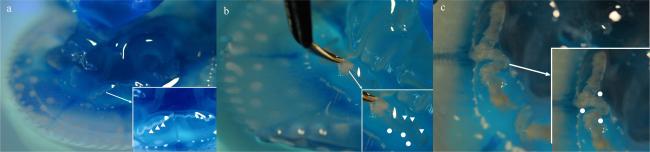
巴布亚硝水母(Mastigias papua)的伞体半球状, 蓝色或黄褐色, 随机分布大小不一的白色斑点, 属刺胞动物门(Cnidarian)钵水母纲(Scyphozoa)根口水母目(Rhizostomeae)硝水母科(Mastigiidae)硝水母属, 广泛分布在太平洋和印度洋, 包括菲律宾、日本、斐济、马来西亚和新加坡等海域(洪惠馨, 2014)。因其观赏价值较高、毒性相对较弱、较为容易饲养等原因, 成为各大海洋馆常见的水母种类之一。巴布亚硝水母的生活史同大多数钵水母相同, 包括水母体阶段的有性生殖和水螅体阶段的无性繁殖, 其水螅体通过性成熟的水母体有性生殖得到, 水母体通过水螅体横裂得到(杨文浩 等, 2022)。
近一个多世纪以来, 有关钵水母的研究除了水域生态环境下的水母暴发之外(Lilley et al, 2009; Marambio et al, 2021), 主要集中在形态特征(Lee et al, 2016; Syazwan et al, 2020)、个体发育(Kienberger et al, 2018)和无性生殖(Adler et al, 2009)等。有性生殖研究中, 大多围绕胚胎发育研究(Dong et al, 2006; Yuan et al, 2008; Schiariti et al, 2012), 关于性别和性腺发育的研究罕有报道。现有少量有关水母性别的研究发现, 除罗盘水母(Chrysaora hysoscella)(Gershwin et al, 2002; Morandini et al, 2010)、咖啡金黄水母(C. melasster) (杨翠华 等, 2012)和安朵仙水母(Cassiopea andromeda) (Hofmann et al, 2002)外, 钵水母纲的水母绝大多数为雌雄异体。
水母的精巢发育一般指精小囊的发育过程。Tiemann等(2010)对冠水母目的紫蓝盖缘水母(Periphylla periphylla)的性腺发育研究发现, 在雄性精巢中可见不同发育时期的精小囊, 且精小囊与生殖上皮一直紧密连接, 早期的精小囊从生殖上皮中生发出来, 发育后期的精小囊通过生殖上皮释放精子到生殖下穴中。旗口水母目和根口水母目的性腺发育过程和冠水母目相似。对海月水母(Aurelia aurita)、狮鬃水母(Cyanea capillata)和Discomedusa lobata的精巢研究发现(Kon et al, 1972; Bratina et al, 1981; Hedwig et al, 2009), 精小囊是精子发生的场所, 是由精巢的生殖上皮细胞侵入至中胶层中形成的。沙海蜇(Nemopilema nomurai)的性腺会随着个体发育即水母体伞径的增加, 精小囊长径也呈指数增长, 在性成熟的雄性个体(伞径超过14cm)中, 有超过8.0×10⁵个处于不同发育时期的精小囊, 以保证其有较长的55d配子释放期(Ohtsu et al, 2007)。Ohtsu等(2007)对沙海蜇精巢发育的研究发现, 早期精巢干瘪并呈乳白色, 精小囊分布十分松散; 随着性腺的发育, 精巢逐渐饱满, 精小囊越来越大彼此间的空隙越来越小; 最后, 成熟的性腺十分饱满, 褶皱明显, 精小囊间几乎无间隙, 精小囊在光照刺激下释放的精子受精率超过90%。
关于水母精子的研究较早, 20世纪70年代, Hinsch等(1973)对刺胞动物门大多数物种的精子进行了超微结构研究, 结果表明海月水母的精子结构与水螅纲水母相似, 精子有细长的头部和较长的鞭毛。刺胞动物的精子为原始型精子, 由短小的头部、略微突出的中段和细长的尾部组成。头部的形态变化较大, 一些水母的精子头部短而圆, 如Nausithoe sp.(Afzelius et al, 1971); 还有一些水母的精子头部细长, 顶部尖锐, 如海月水母(Hinsch et al, 1973)和安朵仙水母(Hinsch, 1974)。Rouse等(2000)对钵水母纲的马赛克水母(Catostylus mosaicus)和澳洲斑点水母(Phyllorhiza punctata)精子的超微结构进行研究发现, 马赛克水母的精子是典型的原始型精子, 有一个短小的头, 一个短圆柱形的核、4个球形的线粒体和一些小的球形电子致密的囊泡组成。由此可见, 原始精子的头部由简单的顶体、球形核、线粒体和自由鞭毛组成, 被认为与体外受精有关。然而目前关于巴布亚硝水母的研究主要集中在水螅体无性生殖和水母体养殖条件的探索等方面(Sugiura, 1965; 杨翠华 等, 2011; 杨文浩 等, 2022), 精巢发育及精子相关的信息都还未有报道。
因此, 本研究以巴布亚硝水母为对象, 对其精巢发育过程外部形态变化以及精小囊形态结构和精子发生的情况进行了观察, 同时, 通过扫描电镜和透射电镜, 研究了精子的超微结构。研究结果不仅是对巴布亚硝水母雄性配子发育过程的记录, 也将成为实现巴布亚硝水母人工繁育的前期重要积累。

1 材料与方法

1.1 实验材料

巴布亚硝水母由来自集美大学水产学院观赏海洋生物协同创新中心的水螅体横裂生殖获得, 根据预实验结果精巢发育实验选择伞径(4.99±0.07)cm(n=12), 精巢未发育的水母。精子超微结构实验选择伞径(7.06±0.11)cm(n=9), 精巢发育成熟的水母。碟状体在圆形旋转式亚克力水母缸(直径20cm, 高度15cm)中养殖, 水母幼体幼体在1t水体的拟克莱斯勒水母养殖系统中养殖, 养殖用水为经紫外灭菌的0.01μm 超滤海水, 盐度(27±1)‰, 水温(25±0.5)℃, 光周期12L:12D, 碟状体每日投喂2次, 水母幼体每日投喂4次, 每次投喂20mL密度为200ind.·mL-1的丰年虫(Artemia sp.)无节幼体, 碟状体喂食30min后吸去残饵, 每3d换水100%。

1.2 巴布亚硝水母精巢发育外部形态观察

将12只性腺未发育的雄性巴布亚硝水母饲养于1t水体的拟克莱斯勒水母养殖系统中, 每3d拍照记录(Nikon D80, 日本)每只水母性腺的外部形态特征, 实验周期为40d。

1.3 光学显微镜下巴布亚硝水母精巢采集与发育观察

每3d采集水母性腺, 采样前用75%的乙醇对采样器械消毒, 并在一次性细胞培养板的孔中加入1mL经0.01μm超滤的天然海水, 用于放置采集的性腺。采样时一手托住水母外伞部, 口腕朝上, 之后将口腕拨到一侧, 用弯头镊子夹取胃丝外周的部分组织并略微上提, 再用手术剪剪下所需性腺组织, 置于细胞培养板中备用。
用吸管将孔板中的性腺组织转移至载玻片上并吸干海水, 用解剖针剥离胃丝并将褶皱的性腺组织展平, 分别在光学倒置显微镜(Leica M80, 德国; Motic MIchrome20, 中国)和体视显微镜(Leica M125C, 德国)下观察, 采用LAS v4.12图形处理软件拍照记录并测量相关参数。

1.4 巴布亚硝水母精巢发育的组织切片制备观察

取1mm×1mm性腺组织, 磷酸缓冲液(phosphate buffer saline, PBS)冲洗后, 放入4%戊二醇溶液中固定过夜; 梯度乙醇脱水; 二甲苯透蜡; 石蜡包埋; 轮转式切片机(Thermo HM325, 美国)连续切片, 厚度为5μm, 在恒温水浴锅(越新HH-2, 中国)中贴片, 二甲苯和梯度乙醇脱蜡, 苏木精-伊红(HE)染色, 再次梯度乙醇脱水后中性树脂封片。正置显微镜(Mshot MF43-N, 中国)观察组织切片, LAS v4.12图形处理软件拍照。

1.5 巴布亚硝水母精子的收集与扫描电镜样品的制备观察

精子束的收集: 熄灯前10min, 将养殖于拟克莱斯勒水母养殖系统中性成熟的雄性个体放到装有1L经紫外灭菌0.01μm超滤天然海水的烧杯中, 在水层表面微充气形成一定水流, 将烧杯水浴在水母原养殖水体中。熄灯1h后将水母从烧杯中取出, 水中可见大量排放的精子束。
精子束的计数: 计数时轻微搅拌1L带有精子束的水体使其混匀, 分别吸取2mL液体于3个一次性细胞培养板的3个孔中, 在倒置显微镜(Leica M80 和 Motic MIchrome 20)下计数精子束数量。
单个精子悬液的制备: 将上述带有大量精子束的烧杯静置30min, 使精子束沉淀于烧杯底部。用吸管吸取烧杯底部海水48mL于24只2mL的离心管中, 离心机(CF1524R, 中国)2000g离心10min, 多次离心弃上清后集中各管沉淀。沉淀加电镜固定液固定; PBS缓冲液漂洗3次, 每次15min; 1%锇酸室温避光固定1.5h; 用移液器将固定液中的精子束混匀并将精子束分散为单个精子悬液。
扫描电镜样品的制备: 取精子悬液置于盖玻片上, 自然晾干; 4%戊二醛和梯度乙醇脱水; 临界点干燥仪(Quorum K850, 英国)干燥处理; 干燥样本紧贴于导电碳膜双面胶上后离子溅射仪(HITACHI MC1000, 日本)喷金30s, 扫描电子显微镜(HITACHI SU8100, 日本)观察拍照。

1.6 巴布亚硝水母精子透射电镜样品制备与观察

实验选择正在排放精子束的巴布亚硝水母, 取300μm×300μm性腺组织, 电镜固定液4℃固定3h; PBS缓冲液漂洗3次, 每次15min; 1%琼脂糖溶液包埋; 梯度乙醇脱水, 每次20min; 100%丙酮2次, 每次15min; 梯度812环氧树脂包埋剂37℃渗透; 烘箱(Bao-80, 中国)37℃过夜; 烘箱60℃聚合48h; 超薄切片机(Leica EM UC7, 德国)60nm超薄切片; 150目方华膜铜网捞片; 2%醋酸铀饱和酒精溶液避光染色8min; 70%乙醇漂洗3次后超纯水漂洗3次; 2.6%枸橼酸铅溶液避二氧化碳染色8min; 超纯水漂洗3次, 室温干燥过夜, 透射电子显微镜(HITACHI HT7800, 日本)下观察拍照。

2 实验结果

2.1 巴布亚硝水母精巢发育外部形态变化

巴布亚硝水母的性腺在伞体中央, 不同发育时期性腺外部形态大不相同。实验开始第1—10d, 巴布亚硝水母精巢外部形态无明显特征, 肉眼仅能看到伞体与口腕连接处外围有一环细丝状的胃丝, 胃丝相连处性腺组织肉眼不可见(图1a)。实验开始第11—25d, 胃丝的外周肉眼可见环状乳白色性腺, 此时的性腺呈梯度发育, 远离胃丝侧发育程度较高, 颜色呈乳白色, 近胃丝侧发育程度较低, 颜色更加透明(图1b)。实验开始第26—40d, 精巢发育成熟, 胃丝的外周明显可见呈浅米黄色的成熟性腺, 性腺呈囊状且褶皱十分明显, 甚至遮掩住胃丝(图1c)。
图1 巴布亚硝水母(Mastigias papua)精巢外部形态变化

a. 实验开始第10d的精巢; b. 第25d的精巢; c.第40d的精巢; a、b和c中右下角为精巢局部放大; 白色箭头所示为局部放大; 白色三角所示为胃丝; 白色圆点所示为精巢

Fig. 1 Testis morphology changes of Mastigias papua.

(a) Testis on the 10th day of the experiment; (b) testis on the 25th day; (c) testis on the 40th day. The lower right corner of (a), (b) and (c) show a partial enlargement of the testis. White arrows show local magnification; white triangles show gastric filaments; white dots show testis
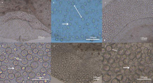
2.2 光学显微镜下巴布亚硝水母精巢发育的形态变化

巴布亚硝水母精巢不同发育时期精小囊形态不同。实验第1—5d, 巴布亚硝水母精巢刚刚开始发育, 生殖上皮较窄, 上面散布着长径(20.61±0.53)μm的透明单层精小囊, 精小囊间隙较大(图 2a、b); 实验第6—7d, 生殖上皮伸长, 精小囊呈梯度发育, 远离胃丝侧的精小囊更大, 长径(35.50±4.19)μm, 精小囊间隙缩小(图2c、d); 实验第8—10d, 生殖上皮进一步伸长, 精小囊也进一步发育变大, 长径(45.89±7.50)μm, 清晰可见不透明的精小囊壁, 精小囊间几乎没有间隙(图2e、f)。实验第11—15d, 性腺梯度发育更加明显, 在远离胃丝侧出现少量不透明的精小囊, 长径(60.13±9.54)μm(图3a、b); 实验第16—25d, 远离胃丝侧基本上全是不透明发育晚期的精小囊, 长径(76.52±7.01)μm, 靠近胃丝侧为透明的早期精小囊(图3c、d)。实验第26—40d, 生殖上皮褶皱十分明显且呈囊状(图4a、b), 上面分布大量发育晚期的精小囊, 长径(96.62±8.69)μm, 内部可见大量头部聚集在中心、尾部朝外的精子束(图4c、d)。
图2 实验第1到10 天巴布亚硝水母(Mastigias papua)的精巢

a、c、e分别为培养不同时间巴布亚硝水母的精巢, b、d、f分别为a、c、e局部放大。双箭头表示精小囊间隙; 箭头所示该阶段典型的精小囊

Fig. 2 The testis of Mastigias papua from 1 to 10d of the experiment.

(a, c, e) The testis of Mastigias papua sampled from different periods, respectively; (b), (d) and (f) are local enlargements of (a), (c) and (e), respectively. The double arrows show the gap of spermatozoa follicles, the arrow shows typical spermatozoa follicles for this phase
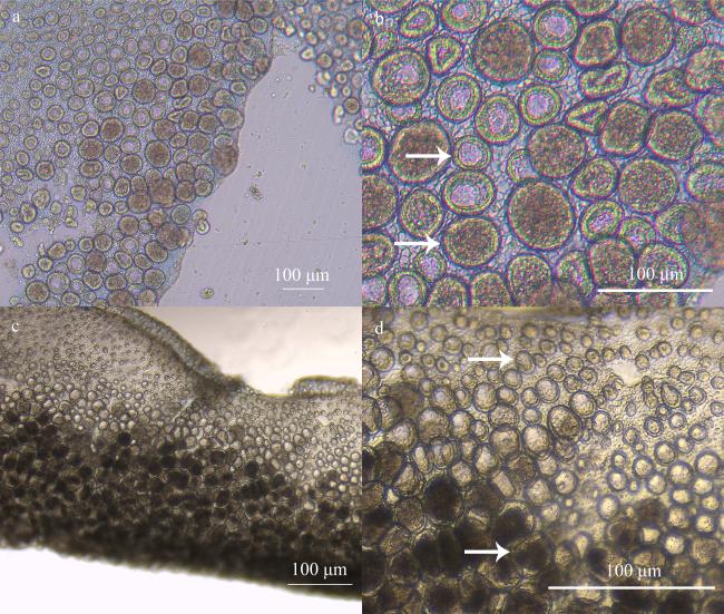
图3 实验第11到25 天巴布亚硝水母(Mastigias papua)的精巢

a和c分别为培养不同时间巴布亚硝水母的精巢, b和d分别为a和c放大的精小囊。箭头所示该阶段典型的精小囊

Fig. 3 The testis of Mastigias papua from 11 to 25d of the experiment.

(a, c) The testis of Mastigias papua sampled from different period, respectively; (b) and (d) are the enlarged spermatozoa follicles of (a) and (c), respectively. The arrows show typical spermatozoa follicles for this phase
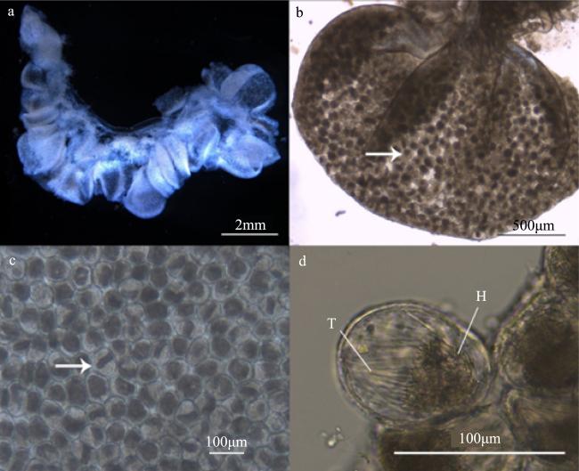
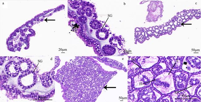
图4 实验第26到40天巴布亚硝水母(Mastigias papua)的精巢

a. 饱满的性腺 b. 囊状精巢 c. 不透明的精小囊 d. 精小囊中精子束。H: 精子头部; T: 精子尾部; 箭头所示处为该阶段性腺典型精小囊

Fig. 4 The testis of Mastigias papua from 26 to 40d of the experiment.

(a) Full gonads; (b) the shape of a sac testis; (c) the opaque spermatozoa follicles; (d) spermatozeugmatas in the spermatozoa follicle. H: head of spermatozoa; T: tail of spermatozoa. The arrows show the typical spermatozoa follicles for this phase

2.3 巴布亚硝水母精巢发育的组织结构变化

巴布亚硝水母不同发育时期的精小囊内部组织结构变化明显。实验第1—10d, 精巢生殖上皮较窄, 精小囊呈卵圆形, 数量较少且分布较为松散, 大多数精小囊较小, 精小囊壁由细胞核大而明显的精原细胞组成, 中央无空腔或空腔很小且染色质松散(图5a、b)。实验第11—25d, 精巢生殖上皮伸长, 依距胃丝距离远近呈梯度发育, 远离胃丝侧的精小囊数量增加且分布更加紧凑, 个体更大且明显可见精小囊壁的细胞染色质固缩, 精小囊腔变大; 靠近胃丝侧的精小囊结构与Ⅰ期精巢中的精小囊相似(图5c、d)。实验第26—40d, 精巢生殖上皮褶皱明显, 精小囊数量最多且分布最为紧凑, 各个精小囊间几乎没有间隙且相互挤压呈不规则的形状, 不同精小囊内精子发生时期不同, 内部可见不同阶段的配子细胞(图5e、f)。
图5 巴布亚硝水母(Mastigias papua)不同阶段精巢组织结构

a、b: 实验开始第1—10d的精巢; c、d: 第11—25d的精巢; e、f: 第26—40d的精巢; H: 精子头部; SG: 精原细胞; SZ: 精子; T: 精子尾部; 粗箭头所示为精巢不同发育时期的精小囊

Fig. 5 Histology of various stages of Mastigias papua testis.

(a, b) The testis from 1 to 10d of the experiment; (c, d) the testis from 11 to 25d of the experiment; (e, f) the testis from 26 to 40d of the experiment. H: head of spermatozoa; SG: spermatogonia; SZ: spermatozoa; T: tail of spermatozoa. The arrows show the spermatozoa follicles at different stages of testis development

图6可见不同时期精小囊内部结构有很大差异。靠近生殖上皮处新分化的精小囊由球状精原细胞组成, 内部无空腔; 随着精巢的发育, 精小囊中央出现空腔, 精原细胞排列更加紧密形成精小囊壁, 在精小囊壁上分化为初级精母细胞进入精小囊腔, 初级精母细胞染色质逐渐固缩, 发育为细胞核更加致密的次级精母细胞, 次级精母细胞进一步发育为精细胞, 精细胞经变态发育为精子, 成熟的精子在精小囊腔中头部聚集, 尾部游离, 聚集成束(图6a)。图6b可见位于生殖下穴边缘的精小囊即将排放精子: 靠近生殖下穴一侧的精小囊壁消失形成排精坑, 精小囊内所有成熟精子以精子束的形式从排精坑处排放至生殖下穴, 后经过口腕排放至水体中, 在实验养殖条件下具有长达3个月的精子排放时间, 且每晚至少排放150 000个精子束。
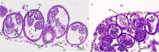
图6 巴布亚硝水母(Mastigias papua)不同发育时期精小囊的组织结构

a. 发育早期的精小囊; b. 发育晚期的精小囊。SG: 精原细胞; S1: 初级精母细胞; S2: 次级精母细胞; SS: 生殖下穴; SP: 排精坑; ST: 精细胞; SZ: 精子; Te: 生殖上皮

Fig. 6 Spermatozoa follicles structure from testis at different developmental stages (Mastigias papua).

(a) Early development of spermatozoa follicles; (b) late development of spermatozoa follicles. SG: spermatogonia; S1: primary spermatocytes; S2: secondary spermatocytes; SZ: spermatozoa; SS: subgenital sinus; SP: spermiation pit; ST: spermatids; Te: testicular epithelium
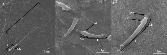
2.4 巴布亚硝水母精子的外部形态

扫描电子显微镜下可见巴布亚硝水母的精子由长尖辣椒状的头部、略微突出的中段和细长的尾部组成(图7)。精子全长为(50.64±3.93)μm (n=12), 长尖辣椒状头部长(7.61±0.63)μm, 顶端尖细, 表面光滑(图7a); 略微突出的中段长(1.00±0.12)μm, 突出部位的宽度为(0.49±0.05)μm, 下方可见明显的锚定装置连接中段和尾部(图7b、c); 尾部长(43.03±4.02)μm, 与头部相比显得十分细长(图7a)。
图7 巴布亚硝水母(Mastigias papua)精子形态

a. 成熟精子完整结构; b、c. 精子头部和中段。Aa: 锚定装置; H: 头部; M: 线粒体; Ms: 中段; T: 尾部

Fig. 7 Mastigias papua spermatozoas morphology.

(a) Complete structure of mature spermatozoa; (b, c) the head and mid-section of mature spermatozoa. Aa: anchoring apparatus; H: head; M: mitochondria; Ms: middle section; T: tail
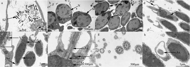
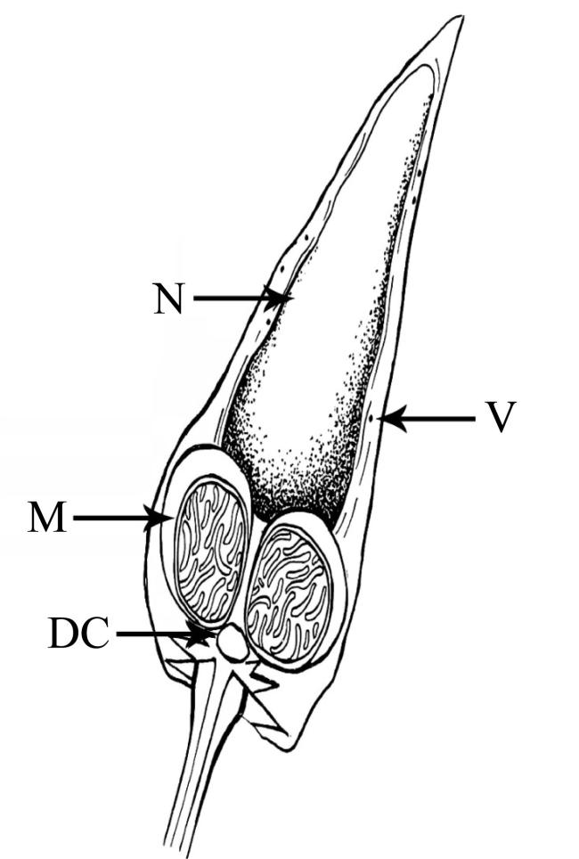
成熟巴布亚硝水母精子头部呈尖辣椒状, 外层为电子致密度较低的质膜, 顶部质膜较为突出无顶体结构, 在质膜中存在一些个体较小的囊泡(图8d、e); 内部为电子致密度较高的细长细胞核, 细胞核较大几乎占满整个头部。细胞核后端出现凹陷, 与中段线粒体相连(图8e)。巴布亚硝水母精子的中段不发达, 由锚定装置和线粒体构成, 锚定装置由远端中心粒和卫星状微管组成, 远端中心粒位于细胞核后端被2个线粒体包围在中间, 卫星状微管与尾部鞭毛的轴丝相连(图8f图9)。尾部由1条细长的鞭毛和2条微管组成, 外部由质膜包裹, 内部轴丝是典型的9+2双联体结构(图8g、h)。
图8 巴布亚硝水母(Mastigias papua)精子的超微结构

a. 性腺中精细胞和成熟精子; b. 精细胞; c. 成熟精子; d—f. 成熟精子头部和中段; g、h. 成熟精子尾部横切图。Ax: 轴丝; DC: 远端中心粒; S: 卫星状微丝; ST: 精细胞; SZ: 精子; PC: 近端中心粒; PM: 质膜; M: 线粒体; Mi: 微管; N: 细胞核; V: 囊泡

Fig. 8 Untrostructure of spermatozoas from Mastigias papua.

(a) Spermatids and mature spermatozoas in the gonad; (b) shows spermatids; (c) shows mature spermatozoas; (d-f) the head and mid-section of mature spermatozoas; (g, h) cross-sections of the tails of mature spermatozoas. Ax: axoneme; DC: distal centriole; S: satellite rays; ST: spermatids; SZ: spermatozoa; PC: proximal centriole; PM: plasma membrane; M: mitochondria; Mi: microtubule; N: nucleus; V: vesicles

图9 巴布亚硝水母(Mastigias papua)精子头部和中段

DC: 远端中心粒; M: 线粒体; N: 细胞核; V: 囊泡

Fig. 9 Head and mid-section structure of Mastigias papua spermatozoa.

DC: distal centriole; M: mitochondria; N: nucleus; V: vesicles

3 讨论

3.1 巴布亚硝水母精巢发育

依据精小囊的形态结构和内部精细胞的成熟情况将巴布亚硝水母精巢发育分为3个时期, 实验第1—10d为Ⅰ期精巢, 实验第11—25d为Ⅱ期精巢, 实验第26—40d为Ⅲ期精巢。不同发育时期特征可见表1
表1 巴布亚硝水母(Mastigias papua)精巢不同时期发育情况

Tab. 1 Development of the testis of Mastigias papua at different stages

精巢分期 外部形态 光学显微镜下 组织切片
Ⅰ期 肉眼不可见(图1a) 精小囊小而透明, 排列松散(图2) 由数个细胞核明显的精原细胞组成的球状精小囊, 内部无空腔(图5a、b)
Ⅱ期 环状乳白色, 由近胃丝处向外性腺透明度降低、颜色加深(图1b) 梯度发育, 由近胃丝处向外精小囊个体变大, 透明度降低(图3) 远离胃丝的精小囊更大, 排列更紧密, 内部有空腔, 可见明显的由精原细胞整齐排列而成的精小囊壁(图5c、d)
Ⅲ期 褶皱呈囊状, 浅米黄色(图1c) 处于不同发育阶段的精小囊, 正在进行精子发生的精小囊占绝大多数(图4) 精小囊内有不同发育阶段的配子细胞, 大量精小囊内充满以精子束存在的成熟精子(图5e、f)
有关水母性腺发育阶段的划分各有不同。Kikinger(1992)依据煎蛋水母(Cotylorhiza tuberculata)是否出现配子将其性腺划分为成熟性腺和未成熟性腺。Lucas等(1998)通过性腺发育指数即性腺和水母体的干重或无灰分干重比表示海月水母的性腺发育程度。此外, 部分文献依据是否存在精小囊和精子是否释放对精巢进行阶段划分(Pitt et al, 2000; Iguchi et al, 2010; Toyokawa et al, 2010)。陈昭廷 等(2017)对海月水母精巢发育的研究中, 依据精小囊和其中生殖细胞的发育阶段将整个性腺发育阶段划分为4个时期: 增殖期、生长期、成熟期和排放期, 但成熟期和排放期的精小囊界限模糊。所以依据巴布亚硝水母精小囊的形态结构松散程度和内部精细胞的成熟情况将其精巢发育分为3个时期。
水母性腺发育不同时期颜色会有一定程度的变化。Schiariti等(2012)对钵水母纲大理石水母(Lychnorhiza lucerna)性腺颜色研究发现, 雌雄水母性腺有暗棕色、亮棕色、白色和蓝绿色, 变化较多。陈昭廷 等(2015)对海月水母精巢发育的研究中表明海月水母的Ⅱ期精巢为灰白色, Ⅳ期则为亮白色或奶白色。实验中巴布亚硝水母不同发育阶段精巢颜色变化不大, Ⅰ期和Ⅱ期为乳白色, Ⅲ期偏米黄色, 这种颜色变化可能与色素、营养物质或生殖细胞数量的积累有关。
钵水母纲的精子发生在精小囊中进行, 结果显示巴布亚硝水母精小囊内有各个发育阶段的精细胞。Schiariti等(2012)对根口水母目大理石水母的精小囊进行组织学研究发现, 不同精小囊内有初级精母细胞、次级精母细胞或精细胞、精子。这种精小囊内有不同发育时期配子细胞的现象在夜光游水母(Pelagia noctiluca)(Sandrini et al, 1991)、海月水母(Lucas et al, 1998)、马赛克水母(Pitt et al, 2000)和沙海蜇(Iguchi et al, 2010)都有报道。巴布亚硝水母不同精小囊内精子发生不同步, 其他水母也存在相同的现象, Schiariti等(2012)表明大理石水母的精巢组织中不同精小囊内的精子发生时期是不同的。Morandini等(2001)对冠水母目的Nausithoe aurea的精巢进行组织学研究发现, 同一精小囊内有多种类型的精细胞且在性腺中不同精小囊发育程度不同。上述结果表明, 水母精巢的不同步发育在钵水母非常普遍。正是这种精小囊内有不同发育时期的精细胞且精巢中各个精小囊发育的不同步性, 使得水母在性成熟期能够长时间排放精子。我们的观测也发现, 巴布亚硝水母在实验条件下培养时具有长达3个月的排精时间, 且每晚至少排放150 000个精子束。

3.2 巴布亚硝水母精子超微结构

巴布亚硝水母为雌雄异体, 精子发生在精小囊内进行, 早期的精原细胞生发于精小囊壁, 之后分化为初级精母细胞、次级精母细胞和精细胞, 靠近生殖下穴一侧的精小囊壁会消失形成排精坑, 精细胞变态为成熟精子后聚集于精小囊一侧, 以精子束的形式从排精坑排放至生殖下穴, 后经过口腕排放。其成熟精子头部边缘的质膜中随机散布电子致密度高的囊泡, 受精时囊泡起到顶体的作用, 释放水解酶溶解卵子外围的放射冠及透明带; 精子鞭毛近端为连接中段和尾部的锚定装置。
Franzén(1967)对刺胞动物精子超微结构的研究发现, 此类动物精子为原始型精子, 包括短而圆的头部, 4~5个线粒体组成的中段和细长的鞭毛, 并认为头部顶端为顶体。然而, Hinsch(1974)对海月水母属(Aurelia)、游水母属(Pelagia)和仙后水母属(Cassiopea)的精子研究发现, 精子头部形态均尖锐细长, 透射电镜发现顶部尖锐处并不存在顶体结构, 取而代之的是一些电子密度较高的囊泡, 并认为该囊泡可起到和顶体一样的作用。与Hinsch(1974)的研究结果相似, 扫描电镜下巴布亚硝水母的精子头部尖锐, 尾部细长; 透射电镜下头部质膜间有电子密度较高的囊泡。Hedwig等(2009)对罗盘水母、桶水母(Rhizostoma pulmo)和海月水母精子的超微结构观察发现, 这些水母精子头部的外形不同但各部分结构差异不大, 头部主要由外围电子密度较低的质膜和内部电子密度较高的细胞核组成, 细胞核形状各异使得头部形态不同。巴布亚硝水母精子头部外围同样是电子密度较低的质膜, 头部形态和电子密度较高的细胞核形态相似, 精细胞的外周质膜内散布有多个体积较小的线粒体, 精子形成过程中, 线粒体移动至细胞核后端融合成2个较大的线粒体, 为精子提供能量(图 8)。Franzén(1996)对水螅纲Cordylophora cmpia的精子超微结构研究表明, 其外部形态和钵水母纲澳洲斑点水母(Rouse et al, 2000)的精子相似, 头部细长且质膜较松散, 中段虽然都有5~6个线粒体, 但钵水母纲的澳洲斑点水母线粒体体积更大, 几乎占满整个中段。巴布亚硝水母和澳洲斑点同属硝水母科(Mastigiidae), 精子外部形态和内部构造十分相似, 头部细长均为尖辣椒状, 质膜中都有电子密度较高的滤泡, 尾部为一条细长的鞭毛, 内部轴丝均具有典型的“9+2”双联体结构(图8d—f)。
Uchida(1926)认为巴布亚硝水母精子头部为短而粗的球形, 但未呈现相关精子形态的照片。本研究扫描电镜表明, 巴布亚硝精子头部为细长的尖辣椒状。Rouse等(2000)对马赛克水母和澳洲斑点水母的精子研究发现, 马赛克水母精子头部为短圆柱形, 澳洲斑点水母精子头部为细长的圆柱形, 二者有不同的形状。钵水母纲中精子形态各异, 夜光游水母的精子头部短粗长度小于4μm, 有一个椭圆的细胞核和球形的线粒体(Franzén, 1967); 狮鬃水母精子头部细长, 为14μm(Arai, 1997); 安朵仙水母精子有一个略微椭圆的头部, 长4μm, 尾部长35μm(Arai, 1997); 煎蛋水母头部圆柱形, 长6μm(Franzén, 1967)。巴布亚硝水母精子头部长(7.61±0.63)μm, 中段长(1.00±0.12)μm, 尾部长(43.03±4.02)μm, 相对于其他水母精子个体较大(表2)。由此可见, 钵水母纲中精子形态和分类地位无明显相关性。
表2 部分钵水母纲精子外部形态

Tab. 2 Scyphozoa spermatozoa morphology

物种 精子头部形态 头部长度/μm 尾部长度/μm 参考文献
夜光游水母 短而粗 <4.00 Franzén(1967)
海月水母 长尖辣椒状 4.15 27.78 Rouse等(2000)
狮鬃水母 细长 14.00 Arai(1997)
马赛克水母 短而粗 2 28.00 Rouse等(2000)
安朵仙水母 略微椭圆 4.00 35.00 Arai(1997)
煎蛋水母 圆柱形 6.00 Franzén(1967)
澳洲斑点水母 细长 6.00 42.00 Rouse等(2000)
巴布亚硝水母 尖辣椒状 7.61±0.63 43.03±4.02 本文章
本研究以巴布亚硝水母为研究对象, 观察记录其精巢外部形态和精小囊发育过程、精子发生过程及精子超微结构, 实验结果可补充巴布亚硝水母精巢发育及精子超微结构的研究, 进一步明确了其精巢发育成熟的时间及过程等, 为其人工有性繁育打下良好基础。

4 结论

巴布亚硝水母在实验条件下(28℃、12L:2D、4次·d-1投喂频率和3000Lux)精巢从开始发育至30d左右成熟, 可持续排放精子束2~3个月。根据性腺外部形态、精小囊结构和精子发生过程, 将其精巢发育阶段分为3个时期, 即Ⅰ期: 精巢外部肉眼不可见, 精小囊小而透明, 排列松散, 内部无空腔; Ⅱ期: 精巢呈环状乳白色, 性腺梯度发育, 远离胃丝的精小囊更大排列更紧密, 内部有空腔; Ⅲ期: 精巢褶皱明显呈囊状米黄色, 光镜下可见处于不同发育阶段的精小囊, 大量精小囊内充满以精子束存在的成熟精子。精小囊梯度成熟且精小囊内存在不同发育阶段的精细胞。巴布亚硝水母的精子发生在精小囊内进行, 精子全长(50.64±3.93)μm, 包括尖辣椒状的头部(7.61±0.63)μm, 略微突出的中段(1.00±0.12)μm和十分细长的尾部(43.03±4.02)μm。头部无顶体, 主要由细胞核和薄的质膜组成, 质膜散布小的囊泡; 中段有两个较大的线粒体, 以锚定装置连接尾部; 尾部轴丝为典型的9+2双联体微管结构。综上所述: 巴布亚硝水母的精巢从开始发育至性成熟分为3个时期, 约30d; 精子较为原始且可分批成熟排放, 有助于延长有性生殖时间, 对物种延续有积极意义。
[1]
陈昭廷, 李琪, 陈四清, 等, 2015. 海月水母精巢发育及排精过程的观察[J]. 动物学杂志, 50(1): 131-140.

CHEN ZHAOTING, LI QI, CHEN SIQING, et al, 2015. The testis development and spermiation process in moon jellyfish[J]. Chinese Journal of Zoology, 50(1): 131-140 (in Chinese with English abstract).

[2]
陈昭廷, 周洋, 顾志峰, 等, 2017. 海月水母(Aurelia aurita)精巢发育及精子的超微结构[J]. 海洋与湖沼, 48(1): 122-129.

CHEN ZHAOTING, ZHOU YANG, GU ZHIFENG, et al, 2017. The testis development and sperm ultrastructure of Aurelia aurita[J]. Oceanologia et Limnologia Sinica, 48(1): 122-129 (in Chinese with English abstract).

[3]
洪惠馨, 2014. 中国海域钵水母生物学及其与人类的关系[M]. 北京: 海洋出版社:60.

HONG HUIXIN, 2014. The relationship between Scyphomedusae biology and human beings in China seas[M]. Beijing: China Ocean Press:60 (in Chinese with English abstract).

[4]
杨翠华, 王玮, 王文章, 2011. 光照和水流对巴布亚硝水母存活时间及存活率的影响[J]. 水产科学, 30(9): 555-558.

YANG CUIHUA, WANG WEI, WANG WENZHANG, 2011. The influences of illumination and water current on survival time and survival rate of spotted jellyfish Mastigias papua[J]. Fisheries Science, 30(9): 555-558 (in Chinese with English abstract).

[5]
杨翠华, 王玮, 王文章, 等, 2012. 咖啡金黄水母的人工培育及生活史观察[J]. 水产科学, 31(12): 708-712.

YANG CUIHUA, WANG WEI, WANG WENZHANG, et al, 2012. Observation of life cycle in brown sea nettle jellyfish Chrysaora melanaster[J]. Fisheries Science, 31(12): 708-712 (in Chinese with English abstract).

[6]
杨文浩, 王淑红, 林子路, 等, 2022. 巴布亚硝水母无性生殖过程及形态变化[J]. 动物学杂志, 57(3): 321-335.

YANG WENHAO, WANG SHUHONG, LIN ZILU, et al, 2022. Asexual reproduction process and morphological changes of Mastigias papus[J]. Chinese Journal of Zoology, 57(3): 321-335 (in Chinese with English abstract).

[7]
ADLER L, JARMS G, 2009. New insights into reproductive traits of scyphozoans: special methods of propagation in Sanderia malayensis GOETTE, 1886 (Pelagiidae, Semaeostomeae) enable establishing a new classification of asexual reproduction in the class Scyphozoa[J]. Marine Biology, 156(7): 1411-1420.

[8]
AFZELIUS B A, FRANZÉN K, 1971. The spermatozoon of the jellyfish Nausithoë[J]. Journal of Ultrastructure Research, 37(1-2): 186-199.

[9]
ARAI M N, 1997. A functional biology of Scyphozoa[M]. London: Chapman & Hall: 137-171.

[10]
BRATINA F, ROTTINI S L, AVIAN M, 1981. Simultaneous presence of Pelagia noctiluca (Forskål) planulae, ephyrae and adults, index of sexual reproduction in the North Adriatic Sea[J]. Bollettino Della Societa Adriaticà di Scienze Naturali in Trieste, 65: 61-68.

[11]
DONG JING, LIU CHUNYANG, WANG YANQING, et al, 2006. Laboratory observations on the life cycle of Cyanea nozakii (Semeostomida, Scyphozoa)[J]. Acta Zoologica Sinica, 52(2): 389-395.

[12]
FRANZÉN Å, 1967. Remarks on spermiogenesis and morphology of the spermatozoon among lower Metazoa[J]. Arkiv För Zoologi, 19(4): 335-342.

[13]
FRANZÉN Å, 1996. Ultrastructure of spermatozoa and spermiogenesis in the hydrozoan Cordylophora caspia with comments on structure and evolution of the sperm in the Cnidaria and the Porifera[J]. Invertebrate Reproduction & Development, 29(1): 19-26.

[14]
GERSHWIN L A, COLLINS A G, 2002. A preliminary phylogeny of Pelagiidae (Cnidaria, Scyphozoa), with new observations of Chrysaora colorata comb. nov[J]. Journal of Natural History, 36(2): 127-148.

[15]
HEDWIG V M, SCHÄFER W, 2009. Vergleichende untersuchungen zur ultrastruktur and zur phylogenetischen bedeutung der spermien der Scyphozoa[J]. Journal of Zoological Systematics and Evolutionary Research, 24(2): 109-122.

[16]
HINSCH G W, CLARK JR W H, 1973. Comparative fine structure of Cnidaria spermatozoa[J]. Biology of Reproduction, 8(1): 62-73.

PMID

[17]
HINSCH G W, 1974. Comparative ultrastructure of cnidarian sperm[J]. American Zoologist, 14(2): 457-465.

[18]
HOFMANN D K, HADFIELD M G, 2002. Hermaphroditism, gonochorism, and asexual reproduction in Cassiopea sp. —an immigrant in the islands of Hawai'i[J]. Invertebrate Reproduction & Development, 41(1-3): 215-221.

[19]
IGUCHI N, LEE H E, YOON W D, et al, 2010. Reproduction of the giant jellyfish, Nemopilema nomurai (Scyphozoa: Rhizostomeae), in 2006-2008 as peripherally-transported populations[J]. Ocean Science Journal, 45(2): 129-138.

[20]
KIENBERGER K, RIERA-BUCH M, SCHÖNEMANN A M, et al, 2018. First description of the life cycle of the jellyfish Rhizostoma luteum (Scyphozoa: Rhizostomeae)[J]. PLoS One, 2018, 13(8): e202093.

[21]
KIKINGER R, 1992. Cotylorhiza tuberculata (Cnidaria: Scyphozoa)-life history of a stationary population[J]. Marine Ecology, 13(4): 333-362.

[22]
KON T, HONMA Y, 1972. Studies on gonad maturity in some marine invertebrates-V. * Seasonal changes in the gonads of the moon jelly[J]. Bulletin of the Japanese Society of Scientific Fisheries, 38(6): 545-553.

[23]
LEE H E, YOON W D, CHAE J, et al, 2016. Re-description of Chrysaora pacifica (Goette, 1886) (Cnidaria, Scyphozoa) from Korean coastal waters: morphology and molecular comparisons[J]. Ocean and Polar Research, 38(4): 295-301.

[24]
LILLEY M K S, HOUGHTON J D R, HAYS G C, 2009. Distribution, extent of inter-annual variability and diet of the bloom-forming jellyfish Rhizostoma in European waters[J]. Journal of the Marine Biological Association of the United Kingdom, 89(1): 39-48.

[25]
LUCAS C H, LAWES S, 1998. Sexual reproduction of the scyphomedusa Aurelia aurita in relation to temperature and variable food supply[J]. Marine Biology, 131(4): 629-638.

[26]
MARAMBIO M, CANEPA A, LÒPEZ L, et al, 2021. Unfolding jellyfish bloom dynamics along the Mediterranean Basin by transnational citizen science initiatives[J]. Diversity, 13(6): 274.

[27]
MORANDINI A C, DA SILVEIRA F L, 2001. Sexual reproduction of Nausithoe aurea (Scyphozoa, Coronatae). Gametogenesis, egg release, embryonic development, and gastrulation[J]. Scientia Marina, 65(2): 139-149.

[28]
MORANDINI A C, MARQUES A C, 2010. Revision of the genus Chrysaora Péron & Lesueur, 1810 (Cnidaria: Scyphozoa)[J]. Zootaxa, 2464(1): 1-97.

[29]
OHTSU K, KAWAHARA M, IKEDA H, et al, 2007. Experimental induction of gonadal maturation and spawning in the giant jellyfish Nemopilema nomurai (Scyphozoa: Rhizostomeae)[J]. Marine Biology, 152(3): 667-676.

[30]
PITT K A, KINGSFORD M J, 2000. Reproductive biology of the edible jellyfish Catostylus mosaicus (Rhizostomeae)[J]. Marine Biology, 137(5-6): 791-799.

[31]
ROUSE G W, PITT K, 2000. Ultrastructure of the sperm of Catostylus mosaicus and Phyllorhiza punctata (Scyphozoa, Cnidaria): Implications for sperm terminology and the inference of reproductive mechanisms[J]. Invertebrate Reproduction & Development, 38(1): 23-34.

[32]
SANDRINI L R, AVIAN M, 1991. Reproduction of Pelagia noctiluca in the central and northern Adriatic Sea[J]. Hydrobiologia, 216- 217(1): 197-202.

[33]
SCHIARITI A, CHRISTIANSEN E, MORANDINI A C, et al, 2012. Reproductive biology of Lychnorhiza lucerna (Cnidaria: Scyphozoa: Rhizostomeae): individual traits related to sexual reproduction[J]. Marine Biology Research, 8(3): 255-264.

[34]
SUGIURA Y, 1965. On the life-history of Rhizostome medusae: III. On the effects of temperature on the strobilation of Mastigias papua[J]. Biological Bulletin, 128(3): 493-496.

[35]
SYAZWAN W M, LOW L B, RIZMAN-IDID M, 2020. First record in Peninsular Malaysia and morphological redescription of Lychnorhiza malayensis (Scyphozoa: Rhizostomeae: Lychnorhizidae)[J]. Raffles Bulletin of Zoology, 68: 32-49.

[36]
TIEMANN H, JARMS G, 2010. Organ-like gonads, complex oocyte formation, and long-term spawning in Periphylla periphylla (Cnidaria, Scyphozoa, Coronatae)[J]. Marine Biology, 157(3): 527-535.

[37]
TOYOKAWA M, SHIMIZU A, SUGIMOTO K, et al, 2010. Seasonal changes in oocyte size and maturity of the giant jellyfish, Nemopilema nomurai[J]. Fisheries Science, 76(1): 55-62.

[38]
UCHIDA T, 1926. The anatomy and development of Rhizostome medusa, Mastigias papua L. Agassiz with observation on the phylogeny of Rhizostomae[J]. Journal of the Faculty of Science University of Tokyo, Section 4, Zoology, 1(4): 45-95.

[39]
YUAN D, NAKANISHI N, JACOBS D K, et al, 2008. Embryonic development and metamorphosis of the scyphozoan Aurelia[J]. Development Genes and Evolution, 218(10): 525-539.

Outlines

/