Exploitation of Marine Resources

Bioactive metabolites from the sponge-derived fungus Penicillium crustosum SCSIO 41442

  • LIANG Jiaqi , 1, 2 ,
  • CHEN Xinqi 1, 2 ,
  • CHEN Yi 1, 2 ,
  • CONG Mengjing 1 ,
  • CAO Qinlin 1 ,
  • HUANG Xiaowen 1, 3 ,
  • LIU Yonghong 1 ,
  • JAYASINGHE Lalith 1, 4 ,
  • CHEN Chunmei , 1 ,
  • ZHOU Xuefeng , 1, 2
Expand
  • 1 CAS Key Laboratory of Tropical Marine Bio-Resources and Ecology, Guangdong Key Laboratory of Marine Materia Medica, South China Sea Institute of Oceanology, Chinese Academy of Sciences, Guangzhou 510301, China
  • 2 University of Chinese Academy of Sciences, Beijing 100049, China
  • 3 Pharmacy school of Guilin Medical University, Guilin 541199, China
  • 4 National Institute of Fundamental Studies, Kandy 20000, Sri Lanka
CHEN Chunmei, email: ;
ZHOU Xuefeng, email:

Received date: 2025-02-18

  Revised date: 2025-03-19

  Online published: 2025-03-25

Supported by

Special Project for Marine Economic Development of Department of Natural Resources of Guangdong Province(GDNRC[2023]37)

Abstract

A new alkaloid, diacedolinate (1), along with fourteen known compounds (2-15) was isolated from the sponge associated fungus Penicillium crustosum SCSIO 41442. The structures of these compounds were determined by spectrum analysis and ECD. All compounds were evaluated for their antioxidant and antimicrobial activities. The results showed that compound 1 exhibited weak antioxidant activity with an IC50 value of (71.00±0.14) μg·mL-1, while compound 2, in contrast, displayed broad antioxidant activity with an IC50 value of (1.25±0.10) μg·mL-1, compared with the positive control, vitamin C. In addition, compounds 9, 10, 11, and 15 demonstrated broad-spectrum antimicrobial activity against a variety of pathogens, including MRSA, Colletotrichum asianum HNM 408, Colletotrichum acutatum HNM RC178, and Alternaria alternate, with MIC values ranging from 2.5 to 160 μg·mL-1. The bioactivities of these compounds are reported here for the first time.

Cite this article

LIANG Jiaqi , CHEN Xinqi , CHEN Yi , CONG Mengjing , CAO Qinlin , HUANG Xiaowen , LIU Yonghong , JAYASINGHE Lalith , CHEN Chunmei , ZHOU Xuefeng . Bioactive metabolites from the sponge-derived fungus Penicillium crustosum SCSIO 41442[J]. Journal of Tropical Oceanography, 2025 , 44(6) : 188 -195 . DOI: 10.11978/2025025

In recent years, antibiotic resistance has emerged as a significant global threat, reducing the effectiveness of curing viral, bacterial, parasitic and fungal infections (Indraningrat et al, 2016). Consequently, the discovery of new bioactive compounds has become very urgent. Marine organisms exhibit substantial differences from their terrestrial counterparts, resulting in distinct metabolic profiles (Carroll et al, 2020). Due to their unique living environments, sponge associated microorganisms are rich in silent genes, which can form a unique physiological metabolism mode and produce secondary metabolites with novel structure and significant activity (Slaby et al, 2017). Furthermore, Penicillium crustosum is an important resource that has yielded diverse classes of bioactive compounds (Kozlovskii et al, 2013), such as alkaloids, diketopiperazines, and polyketides.
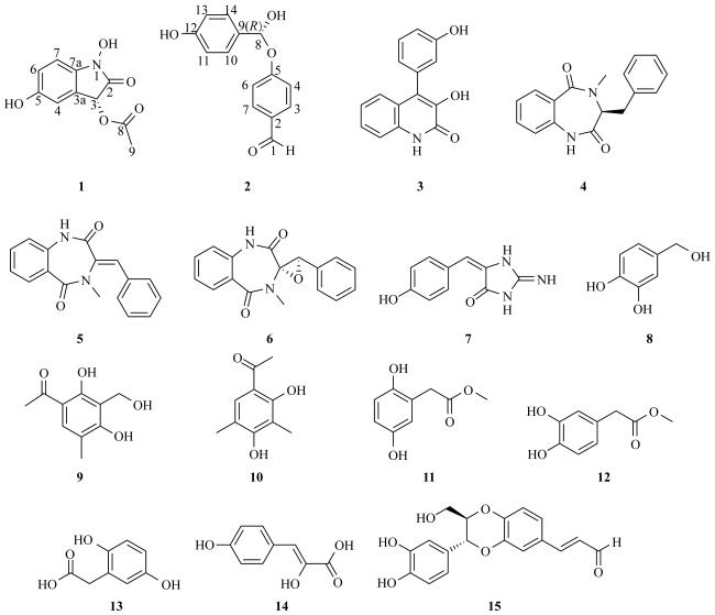
In our search for additional bioactive natural products from marine sponge associated fungi, the strain Penicillium crustosum SCSIO 41442 was isolated from a Callyspongia sp. sponge sample collected near Weizhou Island in the Beibu Gulf of the South China Sea. Chemical investigation of this strain led to a new alkaloid (1), with fourteen other known metabolites (2-15) obtained (Fig. 1). In this paper, we address the isolation, structural elucidation, and biological evaluation of all obtained compounds.
Fig. 1 Chemical structures of compounds 1-15

1 Materials and Methods

1.1 General experimental procedures

NMR data were acquired using a Bruker Avance spectrometer (Bruker, Billerica, MA, USA) operating at 500 and 700 MHz for 1H NMR and 125 and 175 MHz for 13C NMR. High-resolution mass spectrometry was performed on a Bruker TOF-QII mass spectrometer (Bruker, Bille-Rica, MA, USA). Optical rotations were measured on a PerkinElmer MPC 500 polarimeter (Waltham, MA, USA). UV and ECD spectra were recorded on a Chirascan circular dichroism spectrometer (Applied Photophysics, Leatherhead Surrey, United Kingdom). TLC and column chromatography (CC) were performed on silica gel GF254 plates (10-40 μm) and silica gel (200-300 mesh; Qingdao Marine Chemical Factory, Qingdao, China), respectively. Semi-preparative HPLC was conducted using an ODS column (YMC-pack ODS-A, 10 mm × 250 mm, 5 μm). All solvents were of analytical grade. Sea salt was obtained from Guangzhou Haili Aquarium Technology Company (Guangzhou, China).

1.2 Fungal material

The fungus Penicillium crustosum SCSIO 41442 was isolated from a Callyspongia sp. sponge sample collected near Weizhou Island (21.612°N, 108.338°E), Beibu Gulf, South China Sea. The strain was deposited in the Chinese Academy of Sciences Key Laboratory for Tropical Marine Bioresources and Ecology. Based on ITS sequencing, this fungus was identified as Penicillium crustosum (GenBank accession number NR_077153.1).

1.3 Fermentation, extraction and isolation

The fermentation of Penicillium crustosum SCSIO 41442 was performed on rice for 30 d at 26°C. Then the culture was extracted three times with ethyl acetate by ultrasonication, and the organic solvent was evaporated under reduced pressure to obtain the crude extract. The extract was then dissolved in methanol and partitioned with ethyl acetate (1∶1), yielding 100.5 g of the MeOH-soluble fraction.
Nine fractions (P.Fr.1-9) were obtained by fractionation using silica gel CC (200−300 mesh) with an increasing polarity gradient (CH2Cl2/MeOH, from 100∶1 to 0∶1, V/V). ODS CC (Spherical C18, 20−45 μm, 100 A) and MeOH/H2O (V/V, 1∶9−10∶0) were performed on P.Fr.3 (2.4 g) to obtain five subfractions (P.Fr.3.1-3.5). Then, P.Fr.3-2 (216.2 mg), P.Fr.3-3 (154.7 mg) and P.Fr.3-4 (207.5 mg) were purified by semi-preparative HPLC using an ODS column (YMC-pack ODS-A, 10 × 250 mm, 5 µm) to gain 2 (3.0 mg, 45%MeCN/H2O, 2.0 mL·min-1, tR=28.0 min), 4 (3.5 mg, 55%MeOH/H2O, 2.0 mL·min-1, tR=18.0 min), 5 (1.6 mg, 55%MeOH/H2O, 2.0 mL·min-1, tR=20.0 min), and 6 (3.0 mg, 55%MeOH/H2O, 2.0 mL·min-1, tR =12.0 min). ODS CC and MeOH/H2O (1∶9-10∶0, V/V) were performed on P.Fr.4 (2.6 g) to obtain four subfractions (P.Fr.4.1-4.4). Then, P.Fr.4-1 (287.3 mg) and P.Fr.4-3 (246.9 mg) were purified by semi-preparative HPLC to gain 3 (4.5 mg, 55%MeOH/H2O, 3.0 mL·min-1, tR=15.5 min), 7 (3.8 mg, 15%MeOH/H2O, 2.5 mL·min-1, tR=32.0 min), and 14 (1.6 mg, 15%MeOH/H2O, 2.5 mL·min-1, tR=16.8 min). P.Fr.2 (7.9 g), 5 (10.8 g), 6 (2.7 g), and 7 (1.8 g) were purified by HPLC to obtain 1 (3.2 mg, 10%MeCN/H2O, 2.0 mL·min-1, tR=8.0 min), 8 (5.2 mg, 20%MeOH/H2O, 2.5 mL·min-1, tR=9.2 min), 9 (3.1 mg, 45%MeCN/H2O, 2.0 mL·min-1, tR=28.0 min), 10 (33.0 mg, 45%MeCN/H2O, 2.0 mL·min-1, tR=21.0 min), 11 (8.9 mg, 25%MeOH/H2O, 2.0 mL·min-1, tR=19.0 min), 12 (3.5 mg, 25%MeOH/H2O, 2.0 mL·min-1, tR=26.0 min), 13 (27.6 mg, 25%MeOH/ H2O, 2.0 mL·min-1, tR=12.0 min), and 15 (5.0 mg, 25%MeOH/H2O, 2.0 mL·min-1, tR=16.0 min).
Diacedolinate (1) : yellowish oil; [α] -3 (c 0.10, MeOH); UV (MeOH) λmax (log ε) 262 (3.71); 210 (5.13) nm; ECD (0.3mg·mL-1, MeOH) λmax (∆ε) 238 (-3.08); 213 (+4.69) nm; HRESIMS: m/z 224.0555 [M+H]+ (calcd for C10H10NO5 224.0553); for NMR data of 1H and 13C, see Tab. 1.
Tab. 1 13C- and 1H- NMR data of compound 1 in CD3OD (500 MHz, 125 MHz)
Position δH, mult (J in Hz) δC, type
2 174.75, C
3 4.17, s 75.35, CH
3a 133.42, C
4 6.89, d (2.2) 113.05, CH
5 154.52, C
6 6.67, dd (8.3, 2.2) 116.67, CH
7 6.71, d (8.7) 111.76, CH
7a 135.39, C
8 175.73, C
9 2.56, s 30.07, CH3
(R)-4-hydroxy (4-hydroxyphenyl) methoxy) benzaldehyde (2) : Yellow oil; [α] -2 (c 0.10,MeOH); 1H NMR (500 MHz, CD3OD-d4) δH 9.77 (1H, brs, H-1), 6.92 (1H, m, H-3), 7.78 (1H, m, H-4), 7.78 (1H, m, H-6), 6.92 (1H, m, H-7), 4.38 (1H, s, H-8), 7.77 (1H, m, H-10), 6.91 (1H, m, H-11), 6.91 (1H, m, H-13), 7.77 (1H, m, H-14); 13C NMR (125 MHz, CD3OD-d4) δC 192.82 (CHO-1), 130.37 (C-2), 116.56 (CH-3), 130.29 (CH-4), 165.23 (C-5), 130.29 (CH-6), 116.56 (CH-7), 106.14 (CH-8), 130.37 (C-9), 133.43 (CH-10), 116.88 (CH-11), 162.05 (C-12), 116.88 (CH-13), 133.43 (CH-14).
Viridicatol (3): Pink powder; 1H NMR (500 MHz, CD3OD-d4) δH 7.34 (1H, m, H-5), 7.12 (1H, td, J = 8.2, 6.3, 1.4 Hz, H-6), 7.24 (1H, dd, J = 8.4, 1.4 Hz, H-7), 7.35 (1H, m, H-8), 6.81 (1H, m, H-2°), 6.87 (1H, dd, J = 8.4, 2.8 Hz, H-4°), 7.32 (1H, d, J = 7.7 Hz, H-5°), 6.83 (1H, m, H-6°); 13C NMR (125 MHz, CD3OD-d4) δC 160.59 (C-2), 143.21 (C-3), 123.82 (C-4), 123.06 (C-4a), 126.32 (CH-5), 127.97 (CH-6), 122.17 (CH-7), 116.20 (CH-8), 134.33 (C-8a), 136.23 (C-1°), 116.46 (CH-2°), 158.67 (C-3°), 115.95 (CH-4°), 130.56 (CH-5°), 117.93 (CH-6°).
(3S)-1, 4-benzodiazepine-2, 5-diones (4): Yellow oil; [α]-7 (c 0.10, MeOH); 1H NMR (500 MHz, DMSO-d6) δH 10.51 (1H, s, H-1), 4.33 (1H, s, H-3), 7.07 (1H, d, J = 8.0 Hz, H-6), 7.19 (1H, t, J = 7.8 Hz, H-7), 7.85 (1H, t, J = 8.1 Hz, H-8), 7.57 (1H, d, J = 7.6 Hz, H-9), 2.87 (2H, s, H2-10), 7.02 (1H, d, J = 7.2 Hz, H-14), 7.50 (1H, t, J = 7.4 Hz, H-15), 7.72 (1H, t, J = 7.6 Hz, H-16), 7.50 (1H, t, J = 7.4 Hz, H-17), 7.02 (1H, d, J = 7.2 Hz, H-18), 2.95 (3H, s, H3-19); 13C NMR (175 MHz, DMSO-d6) δC 169.22 (C-2), 55.99 (CH-3), 167.67 (C-5), 120.69 (CH-6), 124.10 (CH-7), 136.64 (CH-8), 132.04 (CH-9), 31.49 (CH2-10), 126.99 (C-11), 137.44 (C-12), 130.71 (CH-13), 126.49 (CH-14), 128.41 (CH-15), 129.02 (CH-16), 128.41 (CH-17), 126.49 (CH-18), 28.78 (CH3-19).
7-Hydroxy-3, 10-dehydrocyclopeptine (5): Yellow oil; 1H NMR (700 MHz, CD3OD-d4) δH 7.43 (1H, dd, J = 8.3, 1.3 Hz, H-6), 7.90 (1H, dd, J = 7.9, 1.6 Hz, H-7), 7.36 (1H, dd, J = 7.9, 1.6 Hz, H-8), 7.35 (1H, dd, J = 8.3, 1.3 Hz, H-9), 6.93 (1H, s, H-10), 7.29 (1H, dd, J = 7.9, 1.1 Hz, H-2), 7.41 (1H, t, J = 6.8 Hz, H-3°), 7.54 (1H, ddd, J = 8.1, 7.3, 1.6 Hz, H-4°), 7.38 (1H, t, J = 7.3 Hz, H-5°), 7.13 (1H, dd, J = 8.1, 1.1 Hz, H-6°), 3.12 (3H, s, H3-4-NCH3); 13C NMR (175 MHz, CD3OD-d4) δC 172.40 (C-2), 130.96 (C-3), 36.12 (CH3-4-NCH3), 169.03 (C-5), 133.63 (C-5a), 131.84 (CH-6), 125.90 (CH-7), 134.11 (CH-8), 122.02 (CH-9), 137.95 (C-9a), 131.83 (CH-10), 135.35 (C-1°), 130.18 (CH-2°), 130.24 (CH-3°), 126.75 (CH-4°), 130.24 (CH-5°), 130.18 (CH-6°), 128.41 (CH-17), 126.49 (CH-18), 28.78 (CH3-19).
Cyclopenin (6): Yellow oil; [α]-14 (c 0.10, MeOH); 1H NMR (500 MHz, DMSO-d6) δH 7.23 (1H, dd, J = 6.2, 1.4 Hz, H-4), 7.54 (1H, td, J = 7.9, 7.6, 1.7 Hz, H-5), 7.09 (1H, td, J = 7.6, 7.5, 1.1 Hz, H-6), 6.92 (1H, dd, J = 7.9, 1.6 Hz, H-7), 3.07 (3H, s, H3-10), 6.62 (1H, d, J = 1.6 Hz, H-12), 7.16 (1H, t, J = 7.0 Hz, H-13), 7.30 (1H, t, J = 7.4 Hz, H-14), 7.21 (1H, t, J = 7.2 Hz, H-15), 6.64 (1H, d, J = 2.0 Hz, H-16), 4.36 (1H, s, H-17); 13C NMR (175 MHz, DMSO-d6) δC 165.92 (C-1), 70.15 (C-2), 165.32 (C-3), 131.03 (CH-4), 132.40 (CH-5), 124.22 (CH-6), 121.16 (CH-7), 135.17 (C-8), 127.90 (C-9), 30.84 (CH3-10), 130.90 (C-11), 126.10 (CH-12), 128.72 (CH-13), 130.47 (CH-14), 128.81 (CH-15), 126.42 (CH-16), 63.72 (CH-17).
Hemimycalin D (7): Yellow oil; 1H NMR (500 MHz, DMSO-d6) δH 6.23 (1H, s, H-4), 6.72 (2H, d, J = 9.0 Hz, H2-7, 9), 7.43 (2H, d, J = 9.0 Hz, H2-6, 10); 13C NMR (175 MHz, DMSO-d6) δC 155.40 (C-1), 163.20 (C-2), 126.50 (C-3), 116.50 (CH-4), 123.50 (C-5), 131.60 (CH-6, 10), 114.80 (CH-7, 9), 157.90 (C-8).
3, 4-Dihydroxybenzyl alcohol (8): Black powder; 1H NMR (500 MHz, CD3OD-d4) δH 6.46 (1H, s, H-2), 6.42 (1H, d, J = 8.2 Hz, H-5), 6.53 (1H, d, J = 8.4 Hz, H-6), 3.30 (2H, s, H2-7); 13C NMR (125 MHz, CD3OD-d4) δC 123.62 (C-1), 113.67 (CH-2), 148.67 (C-3), 149.37 (C-4), 116.01 (CH-5), 117.24 (CH-6), 63.09 (CH2-7).
Communal G (9): Yellow powder; 1H NMR (700 MHz, CD3OD-d4) δH 7.60 (1H, s, H-6), 4.68 (2H, s, H2-7), 2.17 (3H, s, H3-8), 2.54 (3H, s, H3-10); 13C NMR (175 MHz, CD3OD-d4) δC 110.92 (C-1), 162.36 (C-2), 117.81 (C-3), 163.52 (C-4), 113.68 (C-5), 133.84 (CH-6), 65.62 (CH2-7), 15.82 (CH3-8), 204.57 (C-9), 26.26 (CH3-10).
Clavatol (10): Yellow powder; 1H NMR (500 MHz, CD3OD-d4) δH 2.53 (3H, s, H3-1), 7.46 (1H, s, H-6°), 2.07 (3H, s, H3-7°), 2.18 (3H, s, H3-8°); 13C NMR (125 MHz, CD3OD-d4) δC 26.19 (CH3-1), 204.4 (C-2), 113.76 (C-1°), 162.16 (C-2°), 111.83 (C-3°),162.33 (C-4°), 117.25 (C-5°), 131.17 (CH-6°), 7.93 (CH3-7°), 16.28 (CH3-8°).
2, 5-Dihydroxy-phenylacetic acid methyl ester (11): Brown powder; 1H NMR (500 MHz, CD3OD-d4) δH 6.62 (1H, d, J = 8.6 Hz, H-3), 6.54 (1H, dd, J = 8.6, 2.9 Hz, H-4), 6.59 (1H, d, J = 2.9 Hz, H-6), 3.55 (2H, s, H2-7), 3.67 (3H, s, H3-9); 13C NMR (125 MHz, CD3OD-d4) δC 123.22 (C-1), 149.56 (C-2), 116.67 (CH-3), 115.67 (CH-4), 151.04 (C-5), 118.56 (CH-6), 36.47 (CH2-7), 174.56 (C-8), 52.33 (CH3-9).
3, 4-Dihydroxyphenylacetic acid methyl ester (12): Brown powder; 1H NMR (500 MHz, CD3OD-d4) δH 6.70 (1H, d, J = 2.1 Hz, H-2), 6.68 (1H, d, J = 8.1 Hz, H-5), 6.56 (1H, dd, J = 8.1, 2.1 Hz, H-6), 3.31 (2H, s, H2-7), 3.66 (3H, s, H3-9); 13C NMR (125 MHz, CD3OD-d4) δC 126.94 (C-1), 116.30 (CH-2), 146.32 (C-3), 145.57 (C-4), 117.34 (CH-5), 121.61 (CH-6), 41.18 (CH2-7), 174.57 (C-8), 52.37 (CH3-9).
Homogentisic acid (13): Brown powder; 1H NMR (500 MHz, CD3OD-d4) δH 6.63 (1H, d, J = 8.6 Hz, H-3), 6.55 (1H, dd, J = 8.6, 2.9 Hz, H-4), 6.62 (1H, d, J = 2.9 Hz, H-6), 3.53 (2H, s, H2-7); 13C NMR (125 MHz, CD3OD-d4) δC 123.58 (C-1), 149.56 (C-2), 116.78 (CH-3), 115.59 (CH-4), 150.98 (C-5), 118.57 (CH-6), 36.75 (CH2-7), 176.21 (C-8).
p-Hydroxyphenylpyruvic acid (14): White powder; 1H NMR (700 MHz, DMSO-d6) δH 6.79 (1H, s, H-3), 7.27 (2H, d, J = 8.6 Hz, H2-5, 9), 6.77 (2H, d, J = 8.6 Hz, H2-6, 8); 13C NMR (175 MHz, DMSO-d6) δC 168.0 (C-1), 138.7 (C-2), 127.2 (CH-3), 124.8 (C-4), 131.0 (CH-5/9), 115.1 (CH-6/8), 156.2 (C, C-7).
Isoamericanin A (15): Brown powder; [α]+2 (c 0.10, MeOH); 1H NMR (500 MHz, CD3OD-d4) δH 6.77 (1H, d, J = 2.1 Hz, H-2), 6.74 (1H, d, J = 7.9 Hz, H-5), 6.72 (1H, dd, J = 8.1, 1.9 Hz, H-6), 4.59 (1H, d, J = 7.6 Hz, H-7), 4.30 (1H, ddd, J = 7.8, 4.7, 2.8 Hz, H-8), 4.23 (1H, dd, J = 12.5, 2.6 Hz, H-9a), 3.38 (1H, dd, J = 12.4, 4.5 Hz, H-9b), 6.87 (1H, d, J = 2.1 Hz, H-2°), 6.80 (1H, d, J = 8.2 Hz, H-5°), 6.92 (1H, dd, J = 8.2, 2.1 Hz, H-6°), 7.31 (1H, d, J = 15.8 Hz, H-7°), 6.66 (1H, dt, J = 15.8, 7.9 Hz, H-8°); 13C NMR (175 MHz, CD3OD-d4) δC 126.41 (C-1), 115.33 (CH-2), 146.29 (C-3), 147.21 (C-4), 116.55 (CH-5), 121.05 (CH-6), 75.58 (CH-7), 75.69 (CH-8), 61.04 (CH2-9), 130.73 (C-1°), 118.88 (CH-2°), 146.21 (C-3°), 147.33 (C-4°), 118.88 (CH-5°), 123.64 (CH-6°), 153.76 (CH-7°), 126.41 (CH-8°), 193.06 (C-9°).


1.4 ECD calculation

The Molecular Merck force field was used to perform the conformational search of compound 1 using Spartan’ 14. Conformers with a Boltzmann population of more than 1% were optimized with Gaussian 09 at the B3LYP/6-31G (d) level in methanol (Cammi et al, 1995). Stable conformers were then chosen for ECD calculations at the B3LYP/6-311G (d, p) level in methanol. The overall ECD data were weighted by Boltzmann distribution, and the ECD curves and enantiomeric ECD curves were produced with a half-band width of 0.33 eV by Guassian view 6.0 software. The UV-corrected Boltzmann calculated contributions for each conformation were used for this purpose.
At the same time, compound 1 conformers whose Boltzmann population is more than 1% are selected for NMR chemical shift calculations. DFT was used to optimize these conformers again at the B3LYP/6-31 G (d) level using GAUSSIAN 09 program. The GIAO method was used to calculate the chemical shift of the NMR data at the PCM/mPW1PW91/6-31 G (d, p) level in methanol. In order to get the final spectrum, we used the Boltzmann distribution theory to average the spectra of the conformers. Unscaled shifts were used to analyze the probability of DP4+. The DP4+ calculations were performed with an Excel spread sheet (sarotti-NMR.weebly.com).

1.5 Antioxidant assay

DPPH radical scavenging activity was tested with minor adjustments to a literature protocol (Sharma et al, 2009). Test compounds and the positive control (vitamin C) were dissolved in methanol to make 2 mg·mL-1 solution and 0.25 mg·mL-1 solution, respectively. Samples with different concentrations (10, 50, 100, 500, 1000 µg·mL-1) were added to 100 μL DPPH (0.2 mmol·L-1). The mixture was stirred vigorously and left in darkness for 30 minutes at room temperature. The absorbance of the resulting solution was measured at 517 nm. Lower absorbance indicates higher free radical scavenging activity, and vice versa for higher absorbance. Vitamin C was used as reference compound and three parallel experiments were designed for each compound. The DPPH radical scavenging activity (%) was calculated as [(Acontrol-Asample)/Acontrol]×100%. Based on the inhibition to the terminal concentration, the inhibitory concentration (IC50), which results in a 50% clearance of the DPPH radical, was estimated.

1.6 Antimicrobial assay

Antimicrobial activities were measured against twelve strains, including Staphylococcus aureus, MRSA, Enterococcus faecalis, Micrococcus luteus, Colletotrichum asianum HNM 408, Colletotrichum acutatum HNM RC178, Colletotrichum gloeosporioides HNM 1003, Pyricularia oryaza HNM 1003, Fusarium oxysporum HNM 1003, Curvularia australiensis, Alternaria alternate and Rhizoctonia solani by the broth microdilution method. Pathogenic and phytopathogenic bacteria were cultivated at 28 ℃ for 24 h and 48 h respectively on a rotating shaker (180 r·min-1). The cultures were diluted with sterile water to achieve an optical absorbance of 0.4~0.6 at 600 nm, before they were transferred into 96-well microtiter plates. Three replicates of each compound were tested in a dilution sequence between 640 and 0.625 μg·mL-1. After 24 hours of culture, the optical absorbance was measured at 600 nm. The lowest concentration which completely inhibited visible growth was recorded in three separate experiments.

2 Results and Discussion

2.1 Structure elucidation

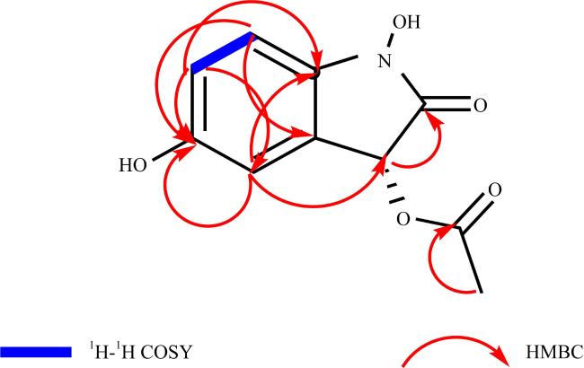
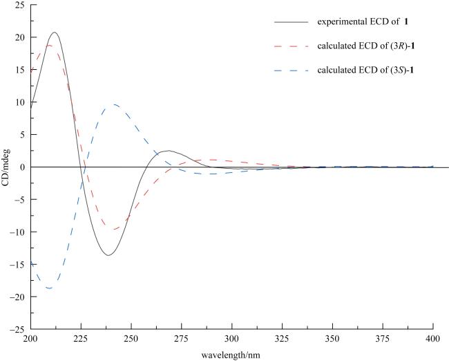
Compound 1 was obtained as a yellow oil with C10H9NO5, which was determined by the HRESIMS peak at m/z 224.0555 [M+H]+ (calcd for C10H10NO5 224.0553), representing 7 degrees of unsaturation. The 1H NMR spectrum (Tab. 1) showed three aromatic proton signals at δ 6.67 (dd, J = 8.3, 2.2 Hz), 6.71 (d, J = 8.7 Hz), and 6.89 (d, J = 2.2 Hz), a signal of oxymethylene at δ 4.17 (s), and a methyl signal at δ 2.56 (s). The 13C NMR (Tab. 1) and HSQC spectra of compound 1 showed 10 carbon signals, including six olefinic carbons at δ 154.52 (C-5), 135.39 (C-7a), 133.42 (C-3a), 116.67 (C-6), 113.05 (C-4), 111.76 (C-7), two carbonyl groups at δ 175.73 (C-8), 174.75 (C-2), an oxygen methylene carbon signal at δ 75.35 (C-3), and a methyl signal at δ 30.07 (9-Me). Based on the unsaturation, compound 1 should contain an additional ring structure apart from the benzene ring. Furthermore, the HMBC correlations of H-4, H-6, H-7/C-5 assigned that the hydroxyl was located at C-5 (Fig. 2). Moreover, in the ECD spectrum, 1 exhibited a positive cotton effect at 213 nm (Δε +4.69), a negative cotton effect at 238 nm (Δε -3.08), and had a shape of curves similar to those of the calculated ECD spectrum of the R-isomer (Fig. 3). These results established the absolute configuration of 1 to be 3R. Therefore, the structure of compound 1 was elucidated as a novel alkaloid, named diacedolinate.
Fig. 2 Key 1H-1H COSY (blue bold lines) and HMBC (red arrows) correlations of compound 1
Fig. 3 Experimental and calculated ECD spectra of compound 1
The other known compounds were identified as (R)-4-hydroxy (4-hydroxyphenyl) methoxy) benzaldehyde (2) (Sulistyowaty et al, 2021), viridicatol (3) (Liu et al, 2021a), (3S)-1, 4-benzodiazepine-2, 5-diones (4) (Mao et al, 2021), 7-hydroxy-3, 10-dehydrocyclopeptine (5) (Liu et al, 2021b), cyclopenin (6) (Liu et al, 2012), hemimycalin D (7) (Shaala et al, 2021), 3, 4-dihydroxybenzyl alcohol (8) (Du et al, 2011), communal G (9) (Newaz et al, 2023), clavatol (10) (Yang et al, 2009), 2, 5-dihydroxyphe-nylacetic acid methyl ester (11) (Dai et al, 1991), 3, 4-dihydroxyphenylacetic acid methyl ester (12) (Li et al, 2019), homogentisic acid (13) (Wang et al, 2016), p-hydroxyphenylpyruvic acid (14) (Bugni et al, 2002), and isoamericanin A (15) (Suzuki et al, 2016), by comparing their NMR data in references.

2.2 Antioxidant and antimicrobial activity

All isolated compounds were evaluated for their antioxidant activity. The results showed that compound 1 had weak antioxidant activity with an IC50 value of (71.00±0.14) μg·mL-1 while compound 2 displayed potent antioxidant activity with an IC50 value of (1.25±0.10) μg·mL-1, compared with the positive control vitamin C (Tab. 2).
Tab. 2 Antioxidant activities of compounds 1 and 2
Compound IC50/( μg·mL-1)
1 71.00±0.14
2 1.25±0.10
Vitamin C a 5.90±0.05

Note: a used as a positive control.

Then, the antimicrobial activities were tested against four pathogenic bacteria, including Staphylococcus aureus, MRSA, Enterococcus faecalis, and Micrococcus luteus, and eight phytopathogenic bacteria, including Colletotrichum asianum HNM 408, Colletotrichum gloeosporioides HNM 1003, Colletotrichum acutatum HNM RC178, Fusarium oxysporum HNM 1003, Pyricularia oryaza HNM 1003, Alternaria alternate, Curvularia australiensis, and Rhizoctonia solani. Among them, compound 9 inhibited Colletotrichum acutatum HNM RC178 with an MIC value of 2.5 μg·mL-1. Clavatol (10) showed broad activity against Colletotrichum asianum HNM 408 with an MIC value of 5.0 μg·mL-1. Compound 11 inhibited MRSA and Colletotrichum asianum HNM 408 with MIC values of 2.5 and 80.0 μg·mL-1, respectively. Compound 15 had weak activity against Alternaria alternate with an MIC value of 160.0 μg·mL-1 (Tab. 3). The other compounds showed no significant activity.
Tab. 3 Antimicrobial activities of compounds 9, 10, 11, and 15
Compound MRSA C. acutatum HNM RC178 C. asianum HNM 408 A. alternate
9 >160 2.5 >160 >160
10 >160 >160 5.0 >160
11 2.5 >160 80.0 >160
15 >160 >160 >160 160.0
Ampicillin a 0.625 - - -
Nystatin b - 0.625 2.5 5.0

Note: a used as a positive control for MRSA; b used as a positive control for phytopathogenic strains.

3 Conclusions

In summary, a new alkaloid, diacedolinate (1), along with 14 known compounds (2-15) were obtained from sponge derived fungus Penicillium crustosum SCSIO 41442. Among them, compounds 2, 4, 5, and 15 were isolated for the first time from sponge associated fungi. In addition, compounds 1 and 2 had antioxidant activity with IC50 values of (71.00±0.14) μg·mL-1and (1.25±0.10) μg·mL-1, respectively. Compounds 9, 10, 11, and 15 showed broad antimicrobial activity against a panel of pathogenic strains with MIC values ranging from 2.5 to 160 μg·mL-1. The biological activities of these compounds were reported for the first time. Therefore, this study enriches the chemical diversity of alkaloids and provides a new perspective for natural product drug development.
[1]
BUGNI T S, CONCEPCIÓN G P, MANGALINDAN G C, et al, 2002. P-Sulfooxyphenylpyruvic acid from the red macro Alga Ceratodictyon spongiosum and its sponge symbiont Haliclona cymaeformis[J]. Phytochemistry, 60(4): 361-363.

DOI

[2]
CAMMI R, TOMASI J, 1995. Remarks on the use of the apparent surface charges (ASC) methods in solvation problems: Iterative versus matrix-inversion procedures and the renormalization of the apparent charges[J]. Journal of Computational Chemistry, 16(12): 1449-1458.

DOI

[3]
CARROLL A R, COPP B R, DAVIS R A, et al, 2020. Marine natural products[J]. Natural Product Reports, 37(2): 175-223.

DOI PMID

[4]
DAI JINRUI, KARDONO L B S, TSAURI S, et al, 1991. Phenylacetic acid derivatives and a thioamide glycoside from Entada phaseoloides[J]. Phytochemistry, 30(11): 3749-3752.

DOI

[5]
DU XIPING, ZHAO BAOBING, ZHENG ZHONGHUI, et al, 2011. Study on the isolation identification and antitumor activity of a phenol derivative from mangrove fungus BYY-1[J]. Journal of Jimei University (Natural Science, 16(6): 424-428.

杜希萍, 赵保兵, 郑忠辉, 等, 2011. 红树内生真菌BYY-1中一个酚类化合物的分离鉴定与抗肿瘤活性[J]. 集美大学学报(自然科学版 ), 16(6): 424-428 (in Chinese with English abstract).

[6]
INDRANINGRAT A A G, SMIDT H, SIPKEMA D, 2016. Bioprospecting sponge-associated microbes for antimicrobial compounds[J]. Marine Drugs, 14(5): 87.

DOI

[7]
KOZLOVSKII A G, ZHELIFONOVA V P, ANTIPOVA T V, 2013. Fungi of the genus Penicillium as producers of physiologically active compounds (Review)[J]. Applied Biochemistry and Microbiology, 49: 1-10.

DOI

[8]
LI HUAXUAN, CUI SHUJUN, XIAO CHAOJIANG, et al, 2019. Phenolic constituents from Ternstroemia gymnanthera and their analgesic activities[J]. Chinese Traditional Patent Medicine, 41(7): 1582-1586.

李骅轩, 崔淑君, 肖朝江, 等, 2019. 厚皮香酚性成分及其镇痛活性[J]. 中成药, 41(7): 1582-1586 (in Chinese with English abstract).

[9]
LIU JUNLIANG, LOU YINGKAI, HU ZHIYU, et al, 2012. Purifications and structure elucidations of cyclopenol and cyclopenin[J]. Journal of Xiamen University (Natural Science, 51(3): 386-390.

刘军亮, 楼盈凯, 胡志钰, 等, 2012. 圆弧菌醇和圆弧菌素的分离纯化及结构解析[J]. 厦门大学学报(自然科学版 ), 51(3): 386-390 (in Chinese with English abstract).

[10]
LIU BILI, QIU YINGKUN, YAN ZHIWEN, et al, 2021a. Studies on marine secondary metabolites of fungus Penicillium polonicum H92[J]. Chinese Journal of Marine Drugs, 40(4): 27-32.

刘碧丽, 丘鹰昆, 颜志文, 等, 2021a. 海洋来源青霉菌Penicillium polonicum H92次级代谢产物的研究[J]. 中国海洋药物, 40(4): 27-32 (in Chinese with English abstract).

[11]
LIU SHUNZHI, HE FENGMING, et al, 2021b. Bioactive compounds derived from the marine-derived fungus MCCC3A00951 and their influenza neuraminidase inhibition activity in vitro and in silico[J]. Natural Product Research, 35(24): 5621-5628.

DOI

[12]
MAO NING, XU WEIFENG, SHAO CHANGLUN, et al, 2021. Research on the alkaloids from the Gorgonian coral-derived fungus Penicillium chrysogenum[J]. Chinese Journal of Marine Drugs, 40(2): 17-20.

毛宁, 徐伟锋, 邵长伦, 等, 2021. 1株柳珊瑚来源真菌Penicillium chrysogenum中生物碱化合物研究[J]. 中国海洋药物, 40(2): 17-20 (in Chinese with English abstract).

[13]
NEWAZ A W, YONG KUO, YI WENWEN, et al, 2023. Antimicrobial metabolites from the Indonesian mangrove sediment-derived fungus Penicillium chrysogenum sp. ZZ1151[J]. Natural Product Research, 37(10): 1702-1708.

DOI

[14]
SHAALA L A, YOUSSEF D T A, 2021. Hemimycalins C-E; cytotoxic and antimicrobial alkaloids with hydantoin and 2-iminoimidazolidin-4-one backbones from the red sea marine sponge Hemimycale sp.[J]. Marine Drugs, 19(12): 691.

DOI

[15]
SHARMA O P, BHAT T K, 2009. DPPH antioxidant assay revisited[J]. Food Chemistry, 113(4): 1202-1205.

DOI

[16]
SLABY B M, HACKL T, HORN H, et al, 2017. Metagenomic binning of a marine sponge microbiome reveals unity in defense but metabolic specialization[J]. The ISME Journal, 11(11): 2465-2478.

DOI

[17]
SULISTYOWATY M I, UYEN N H, SUGANUMA K, et al, 2021. Six new phenylpropanoid derivatives from chemically converted extract of Alpinia galanga (L.) and their antiparasitic activities[J]. Molecules, 26(6): 1756.

DOI

[18]
SUZUKI T, ETO K, KUBOTA Y, et al, 2016. Antioxidative catechol lignans/neolignans isolated from defatted kernel of Jatropha curcas[J]. Journal of Wood Science, 62(4): 339-348.

DOI

[19]
WANG SANLANG, LI H T, ZHANG LIJIE, et al, 2016. Conversion of squid pen to homogentisic acid via Paenibacillus sp. TKU036 and the antioxidant and anti-inflammatory activities of homogentisic acid[J]. Marine Drugs, 14(10): 183.

DOI

[20]
YANG GUOHUA, NENKEP V N, SIWE X N, et al, 2009. An acetophenone derivative, clavatol, and a benzodiazepine alkaloid, circumdatin A, from the marine-derived fungus Cladosporium[J]. Natural Product Sciences, 15(3): 130-133.

Outlines

/