
Journal of Tropical Oceanography ›› 2020, Vol. 39 ›› Issue (6): 93-102.doi: 10.11978/2020014CSTR: 32234.14.2020014
• Marine Biology • Previous Articles Next Articles
Distinct structures of gonads and germ cell development of lined seahorse, Hippocampus erectus
CHEN Lingzhen1,2( ), QIN Geng1,3, WANG Xin1, LIU Yali1, XIAO Wanghong1,2, LIN Qiang1,2,3()
- 1. CAS Key Laboratory of Tropical Marine Bio-resources and Ecology, South China Sea Institute of Oceanology, Chinese Academy of Sciences, Guangzhou 510301, China
2. University of Chinese Academy of Sciences, Beijing 100049, China
3. Southern Marine Science and Engineering Guangdong Laboratory (Guangzhou), Gunagzhou 511458
-
Received:2020-02-02Revised:2020-03-11Online:2020-10-10Published:2020-03-11 -
Contact:LIN Qiang E-mail:chenlingzhen17@mails.ucas.ac.cn;linqiang@scsio.ac.cn -
Supported by:the National Key Research and Development Program(2016YFC1403003);Key Special Project for Introduced Talents Team of Southern Marine Science and Engineering Guangdong Laboratory (Guangzhou)(GML2019ZD0407);Natural Science Foundation of Guangdong Province(2017A030313249)
CLC Number:
- Q953
Cite this article
CHEN Lingzhen, QIN Geng, WANG Xin, LIU Yali, XIAO Wanghong, LIN Qiang. Distinct structures of gonads and germ cell development of lined seahorse, Hippocampus erectus[J].Journal of Tropical Oceanography, 2020, 39(6): 93-102.
share this article
Add to citation manager EndNote|Reference Manager|ProCite|BibTeX|RefWorks
Tab. 1
Morphological characteristics of gonads at different development stages of lined seahorse H.erectus"
| 生长时期/d | 体重/g | 体长/cm | 卵巢 | 精巢 |
|---|---|---|---|---|
| 14 | 0.03±0.01 | 2.20±0.13 | 性腺不可见 | 性腺不可见 |
| 21 | 0.04±0.01 | 2.59±0.10 | 肉眼可见 | 肉眼不可见 |
| 30 | 0.15±0.02 | 3.37±0.74 | 肉眼可见 | 肉眼可见 |
| 40 | 0.33±0.14 | 4.52±0.70 | 性腺细丝状, 透明, 组织切片可辨 | 性腺细丝状, 透明, 组织切片可辨 |
| 55 | 0.41±0.07 | 5.11±0.41 | 肉粉色, 性腺增粗, 细丝状, 肉眼可辨 | 透明, 性腺增长, 细丝状, 肉眼可辨 |
| 70 | 0.55±0.08 | 5.55±0.20 | 肉粉色至浅黄色, 性腺带状, 较精巢略短 | 透明, 性腺增长, 带状 |
| 90 | 2.14±0.73 | 8.36±0.93 | 淡橘色, 性腺增粗, 圆柱状, 较短, 含少量卵黄颗粒 | 乳白色, 性腺增粗增长, 管状, 结缔组织增多 |
| 120 | 4.42±0.80 | 9.75±1.93 | 橘色, 性腺增大, 蛹状, 卵黄颗粒增多, 但不能分离 | 乳白色, 性腺进一步增长、增粗, 管状, 结缔组织增厚 |
| 150 | 5.08±1.74 | 10.01±0.77 | 深橘色, 性腺体积进一步增大, 蛹状, 卵黄颗粒增多, 饱满 | 乳白色, 性腺依然在增粗、增长, 长管状, 结缔组织增厚, 肉眼可见血管 |
| 180 | 9.67±1.28 | 13.58±0.65 | 橘红色, 性腺体积进一步增大, 蛹状, 卵粒饱满, 卵子可从卵巢中挤出 | 乳白色, 性腺圆管状, 较粗, 精液可从精巢中挤出, 肉眼可见血管 |
| 250 | 10.30±0.19 | 13.80±0.22 | 深橘红色, 性腺体积变小, 松弛, 含皱缩空泡, 含卵黄颗粒, 但不能分离 | 乳白色, 性腺圆管状, 较粗, 肉眼可见血管 |
Tab. 1
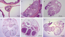
Fig. 1
Histological structures of different stages of H. erectus ovaries. CA: cortical alveoli; DF: degenerated follicle cell; FC: follicle cell; FM: follicle membrane; L: lipid droplet; N: nuclear; Nu: nucleolus; O: oogonia; PO: primary oocyte; VO: vitellogenic oocyte; YM : yolk mass; YS: yolk sphere."
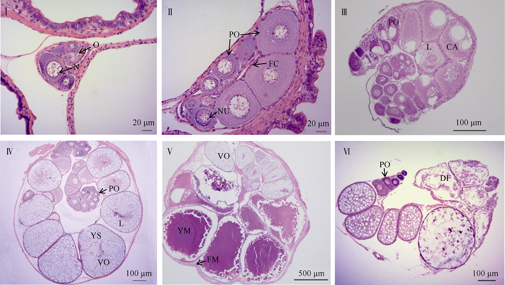
Fig. 1

Fig. 2
Histological and biochemical analysis of different stages of oocytes in H. erectus. (a-e) hematoxylin and eosin staining; (f-j) staining of amino acid; (k-o) staining of fat. BL: basal lamina; CA: cortical alveoli; FC: follicle cell; L: lipid droplet; N: nuclear; Nu: nucleolus; O: oogonia; VE: vitellogenic envelope; YM: yolk mass. Small triangles represent signals of amino acid and fat"
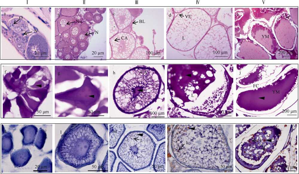
Fig. 2
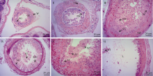
Fig. 3
Histological structures of different stages of H.erectus testis. GE: germinal epithelium; L: lumen; MF: muscle fibres; PSC: primary spermatocyte; SC: sertoli cell; SG: spermatogonia; SSC: secondary spermatocyte; ST: spermatid; SZ: spermatozoa; W: wall"

Fig. 3
| [1] | 陈敬文, 游淑源, 王天娲, 等, 2016. 三种不同脂肪染色方法的比较[J]. 中国组织化学与细胞化学杂志, 25(3):273-276. |
| CHEN JINGWEN, YOU SHUYUAN, WANG TIANWA, et al, 2016. Comparison of three different fat staining methods[J]. Chinese Journal of Histochemistry and Cytochemistry, 25(3):273-276 (in Chinese with English abstract). | |
| [2] | 黄玉喜, 陈平, 张秀梅, 2018. 青岛斋堂岛附近海域薛氏海龙繁殖生物学[J]. 中国水产科学, 25(2):384-394. |
| HUANG YUXI, CHEN PING, ZHANG XIUMEI, 2018. Reproductive biology of Syngnathus schlegeli on the coast of Zhaitang Island[J]. Journal of Fishery Sciences of China, 25(2):384-394 (in Chinese with English abstract). | |
| [3] | 林浩然, 2011. 鱼类生理学[M]. 广州: 中山大学出版社: 292-295. |
| LIN HAORAN, 2011. Fish physiology[M]. Guangzhou: Sun Yat-Sen University Press: 292-295(in Chinese with English abstract). | |
| [4] | 刘晨斌, 徐革锋, 黄天晴, 等, 2019. 鱼类性腺发育研究进展[J]. 水产学杂志, 32(1):46-54. |
| LIU CHENBIN, XU GEFENG, HUANG TIANQING, et al, 2019. A review of research progress on gonadal development in fish[J]. Chinese Journal of Fisheries, 32(1):46-54 (in Chinese with English abstract). | |
| [5] | 刘筠, 1993. 中国养殖鱼类繁殖生理学[M]. 北京: 中国农业出版社: 20-73. |
| LIU JUN, 1993. Propagation physiology of main cultivated fish in China[M]. Beijing: China Agriculture Press: 20-73(in Chinese with English abstract). | |
| [6] | 倪娜, 柳学周, 徐永江, 等, 2011. 条斑星鲽卵巢发育规律和性类固醇激素周年变化研究[J]. 渔业科学进展, 32(3):16-25. |
| NI NA, LIU XUEZHOU, XU YONGJIANG, et al, 2011. The study of gonadal development and steroid hormone annual change in barfin flounder Yerasper moseri[J]. Marine Fisheries Research, 32(3):16-25 (in Chinese with English abstract). | |
| [7] | 施瑔芳, 1988. 鱼类性腺发育研究新进展[J]. 水生生物学报, 12(3):248-258. |
| SHI QUANFANG. Recent advances in the studies on gonad development in fishes[J]. Acta Hydrobiologica Sinica, 12(3):248-258 (in Chinese with English abstract). | |
| [8] | 王晶, 王冰, 李纪同, 等, 2011. 斑马鱼性腺发育的组织学观察[J]. 基因组学与应用生物, 30(2):168-174. |
| WANG JING, WANG BING, LI JITONG, et al, 2011. Histological observation of zebrafish gonad development[J]. Genomics and Applied Biology, 30(2):168-174 (in Chinese with English abstract). | |
| [9] | 徐钢春, 鲍明明, 杜富宽 , 等, 2017. 鱼类性腺发育及产卵类型研究进展[J]. 长江大学学报(自科版), 14(6):43-48. |
| [10] | 尹洪滨, 贾中贺, 姚道霞, 等, 2008. 黄颡鱼性腺分化的组织学观察[J]. 动物学杂志, 43(6):103-108. |
| YIN HONG BIN, JIA ZHONG HE, YAO DAO XIA, et al, 2008. Sex differatiation in pelteobagrus fulvidraco[J]. Chinese Journal of zoology, 43(6):103-108 (in Chinese with English abstract). | |
| [11] | 游秀容, 蔡明夷, 姜永华, 等, 2012. 大黄鱼性腺性别分化的组织学观察[J]. 水产学报, 36(7):1057-1064. |
| YOU XIURONG, CAI MINGYI, JIANG YONGHUA, et al, 2012. Histological observation on gonadal sex differentiation in large yellow croaker (Larimichthys crocea)[J]. Journal of Fisheries of China, 36(7):1057-1064 (in Chinese with English abstract). | |
| [12] | 甄贞, 曲波, 姜毓君, 等, 2014. 中国荷斯坦牛乳腺不同发育时期乳腺细胞内大分子成分变化研究[J]. 东北农业大学学报, 45(4):71-77. |
| ZHEN ZHEN, QU BO, JIANG YUJUN, et al, 2014. Study on changes of macromolecular components during different developmental stages of Chinese Holstein mammary gland[J]. Journal of Northeast Agricultural University, 45(4):71-77 (in Chinese with English abstract). | |
| [13] | 左镇生, 司兆青, 鲁秀红 , 等, 1985. 日本海马卵巢发育规律的初步研究[J]. 水产科学, 4(3):18-21. |
| [14] |
BEGOVAC P C, WALLACE R A , 1988. Stages of oocyte development in the pipefish, Syngnathus scovelli[J]. Journal of Morphology, 197(3):353-369.
pmid: 29895109 |
| [15] | BIAGI F, PIRAS F, FARINA V , et al, 2016. Testis structure, spermatogenesis and sperm morphology in pipefishes of the genus Syngnathus[J]. Acta Zoologica, 97(1):90-101. |
| [16] | CARCUPINO M, BALDACCI A, MAZZINI M , et al, 2002. Functional significance of the male brood pouch in the reproductive strategies of pipefishes and seahorses: a morphological and ultrastructural comparative study on three anatomically different pouches[J]. Journal of Fish Biology, 61(6):1465-1480. |
| [17] | CHEN XIYANG, YI YUNHAI, YOU XINXIN , et al, 2020. High-throughput identification of putative antimicrobial peptides from multi-omics data of the lined seahorse (Hippocampus erectus)[J]. Marine Drugs, 18(1):30. |
| [18] | ELDRIDGE M B, JOSEPH J D, TABERSKI K M , et al, 1983. Lipid and fatty acid composition of the endogenous energy sources of striped bass (Morone saxatilis) eggs[J]. Lipids, 18(8):510-513. |
| [19] | FOSTER J S, VINCENT A C J , 2004. Life history and ecology of seahorses: Implications for conservation and management[J]. Journal of Fish Biology, 65(1):1-61. |
| [20] | FRIEDMAN M , 2004. Applications of the ninhydrin reaction for analysis of amino acids, peptides, and proteins to agricultural and biomedical sciences[J]. Journal of Agricultural and Food Chemistry, 52(3):385-406. |
| [21] | KAMLER E , 2008. Resource allocation in yolk-feeding fish[J]. Reviews in Fish Biology and Fisheries, 18(2):143-200. |
| [22] | KVARNEMO C, SIMMONS L W , 2004. Testes investment and spawning mode in pipefishes and seahorses (Syngnathidae)[J]. Biological Journal of the Linnean Society, 83(3):369-376. |
| [23] | LIN QIANG, LIN JUNDA, LU JUNYI , et al, 2008. Biochemical composition of six seahorse species, Hippocampus sp., from the Chinese Coast[J]. Journal of the World Aquaculture Society, 39(2):225-234. |
| [24] | LIN QIANG, LI GANG, QIN GENG , et al, 2012. The dynamics of reproductive rate, off spring survivorship and growth in the lined seahorse, Hippocampus erectus Perry, 1810[J]. Biology Open, 1(4):391-396. |
| [25] | LIN QIANG, FAN SHAOHUA, ZHANG YANHONG , et al, 2016. The seahorse genome and the evolution of its specialized morphology[J]. Nature, 540(7633):395-399. |
| [26] |
LUBZENS E, YOUNG G, BOBE J , et al, 2010. Oogenesis in teleosts: how fish eggs are formed[J]. General & Comparative Endocrinology, 165(3):367-389.
pmid: 19505465 |
| [27] | NAGAHAMA Y, HIROSE K, YOUNG G , et al, 1983. Relative in vitro effectiveness of 17α, 20β-dihydroxy-4-pregnen-3-one and other pregnene derivatives on germinal vesicle breakdown in oocytes of ayu (Plecoglossus altivelis), amago salmon (Oncorhynchus rhodurus), rainbow trout (Salmo gairdneri) and goldfish (Carassius autatus)[J]. General and Comparative Endocrinology, 51(1):15-23. |
| [28] | NAKAMURA M, KOBAYASHI T, CHANG X T , et al, 1998. Gonadal sex differentiation in teleost fish[J]. Journal of Experimental Zoology, 281(5):362-372. |
| [29] | NEJEDLI S, PETRINEC Z, KUŽIR S , et al, 2004. Annual oscillation of ovarian morphology in european pilchard (Sardina pilchardus Walbaum) in the northern adriatic sea[J]. Veterinarski Arhiv, 74(2):97-106. |
| [30] | NISHIMURA T, TANAKA M , 2014. Gonadal development in fish[J]. Sexual Development, 8(5):252-261. |
| [31] | NOVELLI B, SOCORRO J A, CABALLERO M J , et al, 2015. Development of seahorse (Hippocampus reidi, Ginsburg 1933): histological and histochemical study[J]. Fish Physiology and Biochemistry, 41(5):1233-1251. |
| [32] | OFELIO C, DÍAZ A O, RADAELLI G , et al, 2018. Histological development of the long-snouted seahorse Hippocampus guttulatus during ontogeny[J]. Journal of Fish Biology, 93(1):72-87. |
| [33] | ORBAN L, SREENIVASAN R, OLSSON P E , 2009. Long and winding roads: testis differentiation in zebrafish[J]. Molecular and Cellular Endocrinology, 312(1-2):35-41. |
| [34] |
PARENTI L R, GRIER H J , 2004. Evolution and Phylogeny of Gonad Morphology in Bony Fishes[J]. Integrative and Comparative Biology, 44(5):333-348.
doi: 10.1093/icb/44.5.333 |
| [35] | PIRAS F, BIAGI F, TADDEI A R , et al, 2016. Male gonads morphology, spermatogenesis and sperm ultrastructure of the seahorse Hippocampus guttulatus (Syngnathidae)[J]. Acta Zoologica, 97(3):325-333. |
| [36] |
PLANAS M, QUINTAS P, CHAMORRO A , et al, 2010. Female maturation, egg characteristics and fatty acids profile in the seahorse Hippocampus guttulatus[J]. Animal Reproduction Science, 122(1-2):66-73.
doi: 10.1016/j.anireprosci.2010.07.008 pmid: 20727689 |
| [37] |
SAAVEDRA M, MASDEU M, HALE P , et al, 2014. Dietary fatty acid enrichment increases egg size and quality of yellow seahorse Hippocampus kuda[J]. Animal Reproduction Science, 145(1-2):54-61.
doi: 10.1016/j.anireprosci.2013.08.004 |
| [38] | SAAVEDRA M, BATISTA H, POUSÃO-FERREIRA P , 2016. Dietary fatty acid enrichment during the spawning season increases egg viability and quality in Hippocampus hippocampus[J]. Aquaculture Nutrition, 22:343-351. |
| [39] | SELMAN K, WALLACE R A , 1989. Cellular aspects of oocyte growth in Teleosts[J]. Zoological Science, 6:211-231. |
| [40] | SELMAN K, WALLACE R A, PLAYER D , 1991. Ovary of the seahorse, Hippocampus erectus[J]. Journal of Morphology, 209(3):285-304. |
| [41] | SELMAN K, WALLACE R A, SARKA A , et al, 1993. Stages of oocyte development in the zebrafish, Brachydanio rerio[J]. Journal of Morphology, 218:203-224. |
| [42] | SILVEIRA R B , 2005. Dinâmica populacional do cavalo-marinho Hippocampus reidi no manguezal de Maracaípe, Ipojuca, Pernambuco, Brasil[D]. Brasil: Universidade Católica do Rio Grande do Sul: 1-111. |
| [43] | SOGABE A, TAKATA H, KOBAYASHI Y , 2013. Ovarian structure and mode of egg production in the seaweed pipefish Syngnathus schlegeli (Syngnathidae)[J]. Ichthyological Research, 60(1):85-88. |
| [44] |
STÖLTING K N, WILSON A B , 2007. Male pregnancy in seahorses and pipefish: beyond the mammalian model[J]. BioEssays, 29(9):884-896.
pmid: 17691105 |
| [45] | STRÜSSMANN C A, NAKAMURA M , 2002. Morphology, endocrinology, and environmental modulation of gonadal sex differentiation in teleost fishes[J]. Fish Physiology and Biochemistry, 26(1):13-29. |
| [46] |
TAKAHASHI T, FUJIMORI C, HAGIWARA A , et al, 2013. Recent advances in the understanding of teleost medaka ovulation: the roles of proteases and prostaglandins[J]. Zoological Science, 30(4):239-247.
doi: 10.2108/zsj.30.239 |
| [47] |
UCHIDA D, YAMASHIT M, KITANO T , et al, 2002. Oocyte apoptosis during the transition from ovary-like tissue to testes during sex differentiation of juvenile zebrafish[J]. The Journal of Experimental Biology, 205:711-718.
pmid: 11914381 |
| [48] | VAN LOOK K J W, DZYUBA B, CLIFFE A , et al, 2007. Dimorphic sperm and the unlikely route to fertilisation in the yellow seahorse[J]. Journal of Experimental Biology, 210(3):432-437. |
| [49] | WHITTINGTON C M, GRIFFITH O W, QI WEIHONG , et al, 2015. Seahorse brood pouch transcriptome reveals common genes associated with vertebrate pregnancy[J]. Molecular Biology & Evolution, 32(12):3114-3131. |
| [50] | XU HONGYAN, LI MINGYOU, GUI JIANFANG , et al, 2010. Fish germ cells[J]. Science China Life Sciences, 53(4):435-446. |
| [1] | WU Tao, PAN Ying, LIU Yiming, LIAN Changpeng, XU Bingjie, WANG Chaoqi, YANG Ling. Testis development, spermatogenesis and sperm ultrastructure of Lutraria sieboldii in the Beibu Gulf, Guangxi [J]. Journal of Tropical Oceanography, 2024, 43(2): 69-80. |
| [2] | LIAN Changpeng, WU Tao, WANG Chaoqi, YANG Ling, PAN Ying. Ovarian development, histology of oogenesis and yolk formation of Tapes conspersus from the Beihai Yingpan, Guangxi [J]. Journal of Tropical Oceanography, 2022, 41(5): 170-179. |
| [3] | MA De-you, YOU Feng, WEN Ai-yun, TAN Xun-gang, XU Yong-li, ZHANG Li-jing, ZHANG Pei-jun. A comparison of gonad development between laboratory-reared and wild Branchiostoma japonicum [J]. Journal of Tropical Oceanography, 2013, 32(4): 78-83. |
| [4] | HUANG Jian-hua,YANG Qi-bin,LIN Hei-zhao,CHEN Xu,ZHOU Fa-lin,WEN Wei-geng,J. Fatty acid composition of wild female Penaeus monodon broodstock from the northern South China Sea during ovarian development [J]. Journal of Tropical Oceanography, 2011, 30(1): 144-151. |
| [5] |
ZOU Zhi-hua,ZHANG Zi-ping,WANG Yi-lei,CHEN Jin-min,JIA Xi-wei,WANG Shu-hong,LIN Peng . The construction of normalized cDNA library and preliminary EST analysis from the gonad development and sexual differentiation related organs of Scylla serrata [J]. Journal of Tropical Oceanography, 2009, 28(6): 88-94. |
| [6] | CAO Fu-jun. Observation on the ovary development of Scartelaos virids [J]. Journal of Tropical Oceanography, 2009, 28(6): 123-129. |
|
||