
Journal of Tropical Oceanography ›› 2024, Vol. 43 ›› Issue (2): 59-68.doi: 10.11978/2023090CSTR: 32234.14.2023090
• Marine Biology • Previous Articles Next Articles
Spermary development, spermatogenesis and sperm ultrastructure of Paphia textile in the Beibu Gulf, Guangxi
XU Bingjie1,2( ), LIU Yiming1,2, XING Qinggan1,2, LIAN Changpeng1,2, WU Tao1,2, PAN Ying1,2()
- 1. College of Animal Science and Technology, Guangxi University, Nanning 530004, China
2. Key Laboratory of Aquatic Healthy Breeding and Nutrition Regulation of Guangxi Universities, Nanning 530004, China
-
Received:2023-06-30Revised:2023-08-22Online:2024-03-10Published:2024-03-26 -
Supported by:Guangxi Innovation Driven Development Project(Guike AA19254032)
Cite this article
XU Bingjie, LIU Yiming, XING Qinggan, LIAN Changpeng, WU Tao, PAN Ying. Spermary development, spermatogenesis and sperm ultrastructure of Paphia textile in the Beibu Gulf, Guangxi[J].Journal of Tropical Oceanography, 2024, 43(2): 59-68.
share this article
Add to citation manager EndNote|Reference Manager|ProCite|BibTeX|RefWorks
Tab.1
Morphological parameters of P. textile"
| 参数 | 壳长/mm | 壳宽 /mm | 壳高/mm | 体质量/g | 软体质量/g |
|---|---|---|---|---|---|
| 平均值±标准差 | 62.73±5.39 | 23.45±2.62 | 36.90±3.41 | 40.05±11.90 | 8.59±2.99 |
| 最大值 | 73.97 | 27.03 | 42.05 | 67.89 | 11.14 |
| 最小值 | 52.10 | 16.60 | 29.32 | 20.45 | 4.16 |
Tab.1
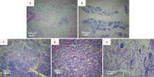
Fig. 1
Histological section of different spermary development stages in P. textile. (a) Proliferating stage; (b) Growing stage; (c) Maturing stage; (d) Spawning stage; (e) Resting stage"
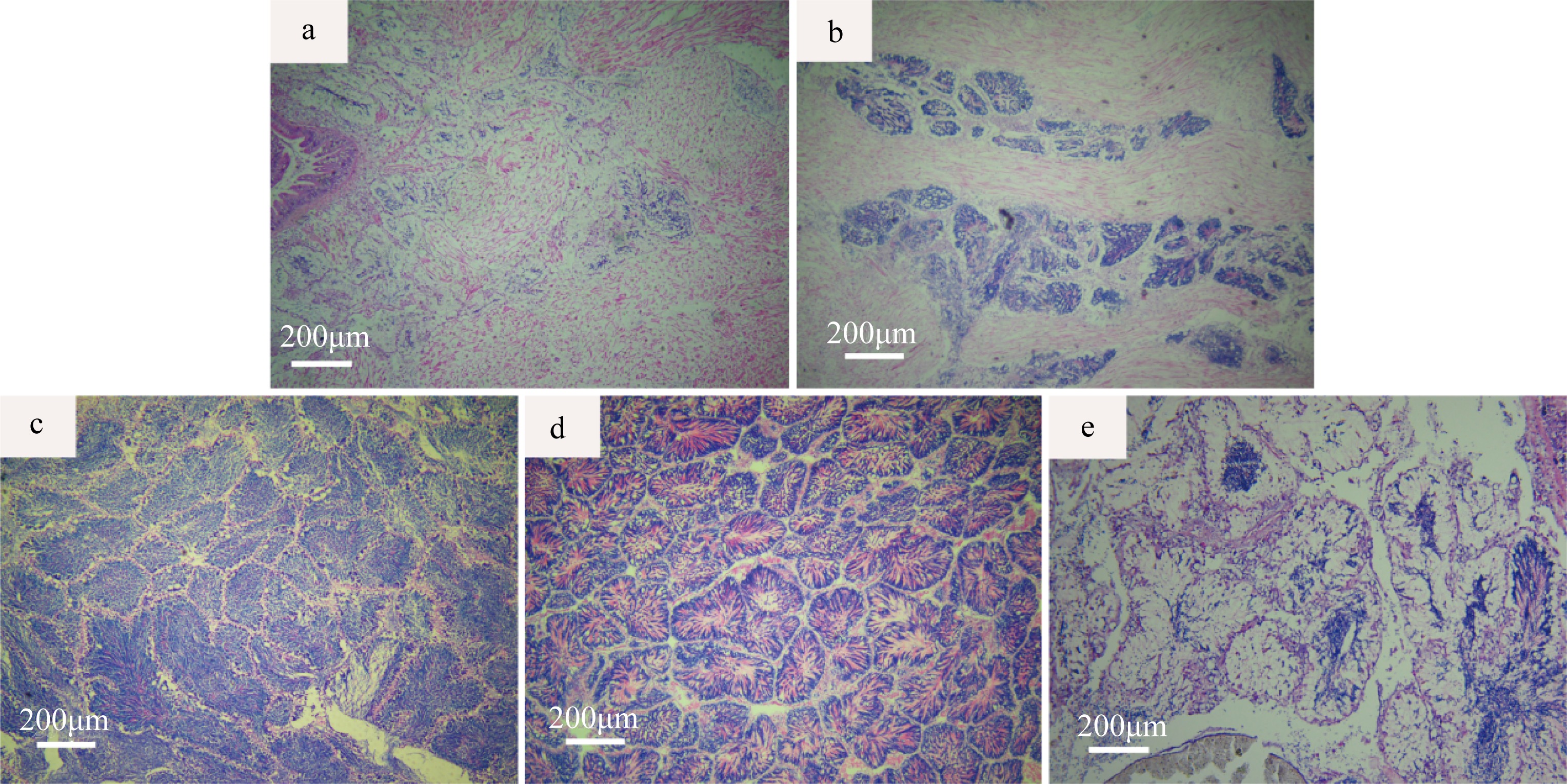
Fig. 1
Tab. 2
Annual distribution of spermary development stages of P. textile in the Beibu Gulf, Guangxi"
| 取样日期 | 肥满度 | 精巢发育分期 | 总数/个 | 盐度/‰ | 水温/℃ | ||||
|---|---|---|---|---|---|---|---|---|---|
| Ⅰ | Ⅱ | Ⅲ | Ⅳ | Ⅴ | |||||
| 2020-11-29 | 25.20 | 8 | 5 | 1 | 14 | 27.4 | 22.0 | ||
| 2020-12-21 | 21.09 | 7 | 2 | 9 | 27.5 | 17.2 | |||
| 2021-01-08 | 18.33 | 1 | 5 | 2 | 8 | 29.4 | 15.6 | ||
| 2021-02-24 | 16.64 | 2 | 8 | 10 | 30.9 | 17.3 | |||
| 2021-03-18 | 20.26 | 11 | 11 | 30.0 | 21.0 | ||||
| 2021-04-22 | 21.24 | 8 | 8 | 30.0 | 27.5 | ||||
| 2021-05-20 | 27.90 | 7 | 2 | 4 | 13 | 28.0 | 29.5 | ||
| 2021-06-23 | 28.24 | 3 | 7 | 1 | 10 | 28.4 | 30.5 | ||
| 2021-07-15 | 34.89 | 1 | 4 | 2 | 7 | 29.0 | 29.7 | ||
| 2021-08-18 | 34.83 | 2 | 6 | 2 | 10 | 30.0 | 29.8 | ||
| 2021-09-17 | 37.40 | 2 | 6 | 8 | 31.0 | 29.8 | |||
| 2021-10-20 | 33.92 | 4 | 2 | 6 | 29.0 | 25.5 | |||
Tab. 2
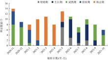
Fig. 2
Staged distribution of the annual spermary development in P. textile"
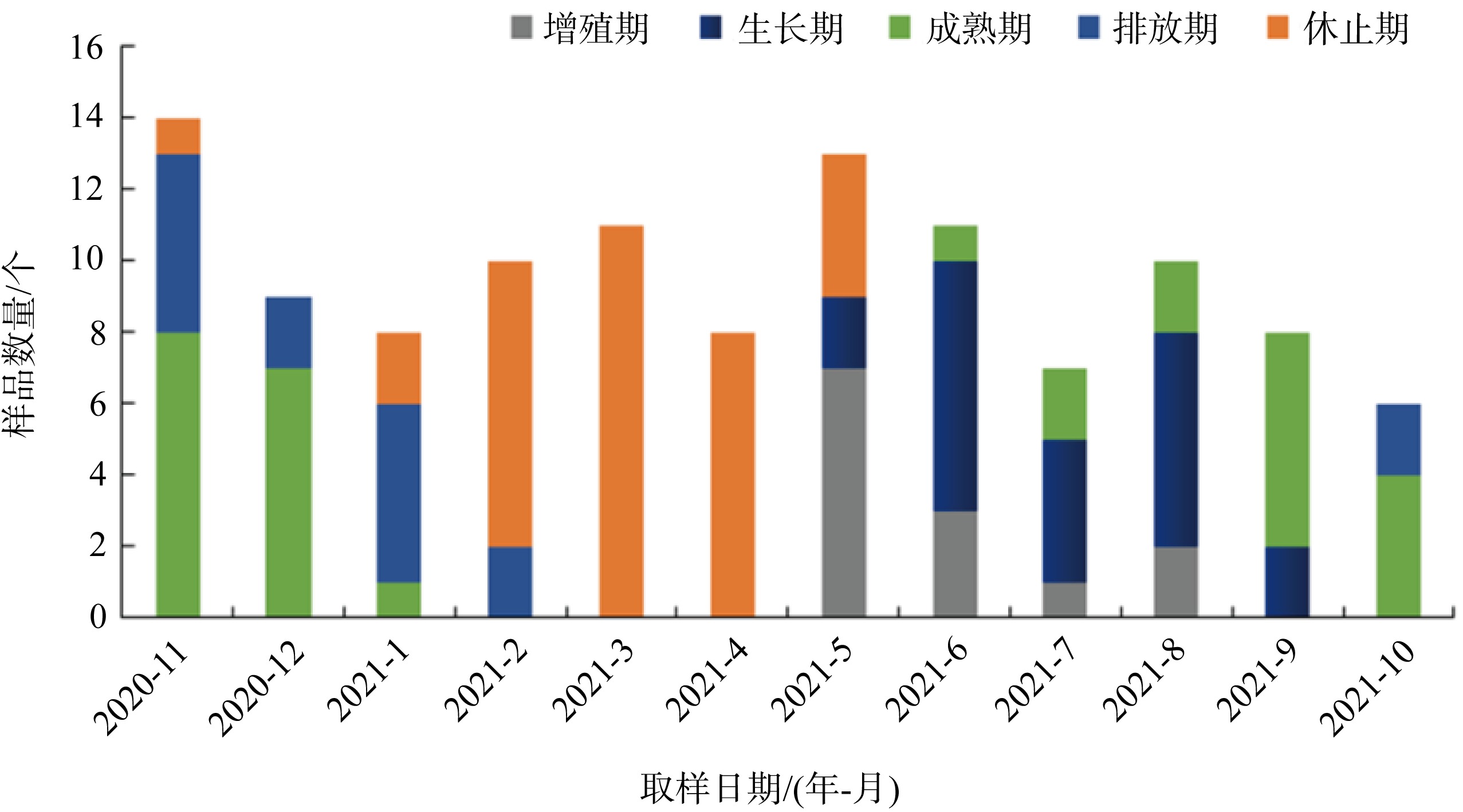
Fig. 2
Tab. 3
Development characteristics of the spermatid of P. textile at various stages"
| 发育分期 | 出现时期及位置 | 细胞特点 |
|---|---|---|
| 精原细胞 | 精巢发育各个阶段均可观察到精原细胞, 增殖期较多。由滤泡壁上的上皮细胞增殖而来, 分布在滤泡基膜和生精小管附近。可分为 A型和B型精原细胞。A型细胞一般贴附在滤泡壁或者基膜上, B型分布在滤泡各个部位或紧贴A型精原细胞。 | 精原细胞形状为圆形或椭圆形, 体积在精子发育过程中最大。A型精原细胞胞内物质少、核仁显现, 但不位于细胞中央, 直径在5.04~6.43μm。B型胞内物质趋于紧密、核仁不明显, 直径在4.89~6.98μm。此期精原细胞染色较浅。 |
| 生长初级精母细胞 | 在精巢发育的生长期较多, 成熟期也存在。生长初级精母细胞由精原细胞A型分化而来, 主要分布在生精小管附近, 靠近滤泡壁。 | 形状为椭圆形或者圆形, 直径在3.56~4.78μm, 比精原细胞小, 细胞核不位于细胞的中央。 |
| 生长次级精母细胞 | 生长期和成熟期数量最多, 分布不规律, 在生精小管附近。 | 形状为椭圆形或者圆形, 细胞直径比初级精母细胞小, 直径在2.12~3.87μm, 此期细胞的核膜、核仁不清晰, 细胞核染色质开始浓缩。 |
| 精细胞 | 成熟期、排放期可观察到较多的精细胞, 分布在滤泡中生精小管周围。 | 精细胞分为前期、中期、后期3个时期。前期主要为椭圆形或者圆形, 至中期大部分转变为椭圆形, 后期为成熟精子。此期染色质高度浓缩, 染色程度高。 |
| 成熟精子 | 在成熟期和排放期成熟精子数量最多, 分布在生精小管上, 以头部朝向滤泡壁、尾部聚集成束朝向滤泡腔中心。 | 精细胞经过复杂变化形成具有特殊结构的精子。成熟精子呈子弹头形状, 头部粗、尾部细。染色质高度浓缩, 染色程度深。在滤泡中呈菊花状散布, 聚集成束。 |
Tab. 3
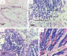
Fig. 3
The different stages of male reproductive cell of P. textile. (a) Spermatogonium phase, Spe-A: spermatogonia A, Spe-B: spermatogonia B; (b) Primary spermatocyte, Ps: Primary spermatocyte, Ss: Secondary spermatocyte; (c) Secondary spermatocyte phase, Ps: Primary spermatocyte, Ss: Secondary spermatocyte; (d) Sperm cells period and sperm phase, St: Seminiferous tubule, Sip: Spermatid in prophase, Sim; Spermatid in metaphase, Ms: Mature Sperm"
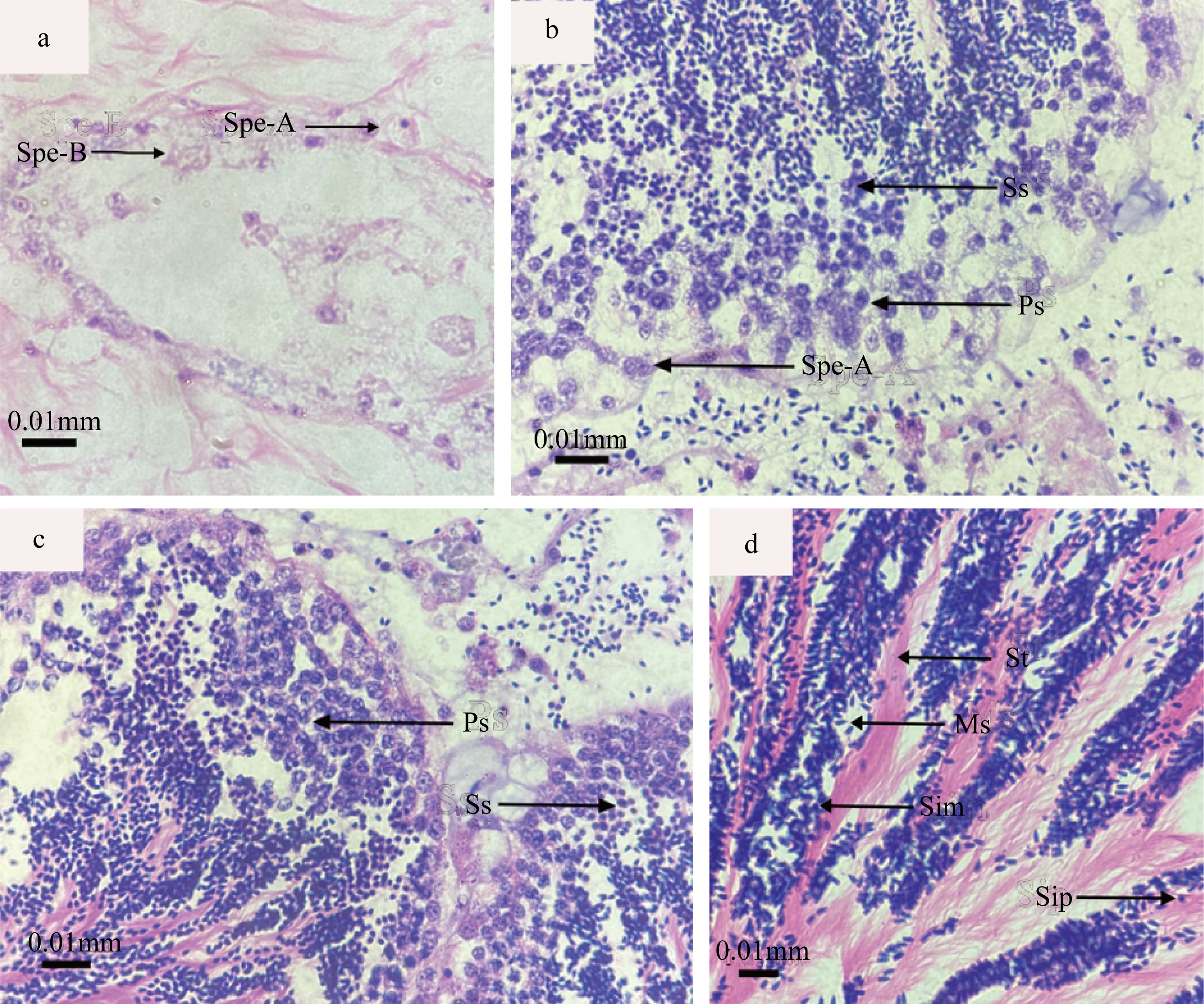
Fig. 3

Fig. 4
Ultrastructural observations on the sperm of P. textile. (a) Sperm, showing head (H); (b) Sperm head and mid-section, showing acrosome (A), nucleus (N), mitochondria (M), flagellum (F); (c) Cross section of mid-section, showing distal centrioles (DC) and mitochondria (M); (d) Sperm longitudinal section, showing plasma membrane (PM), front nuclear pocket (FP), implantation fossa (IF); (e) Longitudinal section of mid-section, showing subacrosomal space (SS), posterior pit (PP), nuclear membranes (NM); (f) Mid-sperm cross section, showing proximal centrioles (PC), nucleus (N); (g) Longitudinal section of tail flagella, showing flagellum (F), axoneme (AX); (h) Longitudinal section of tail flagella, showing central microtubule (CM), doublet microtubules (DM), plasm membrane (PM); (i) Cross section of tail flagella, showing flagellum (F); (j) Cross section of tail flagella, showing microtubule (CM), doublet microtubules (DM)"

Fig. 4
Tab. 4
Ultrastructural comparison of the sperm of different marine bivalve shellfish"
| 物种 | 精子长度/μm | 顶体形状 | 精核长宽比值 | 线粒体数目 | 尾部结构 | 参考文献 | |
|---|---|---|---|---|---|---|---|
| 织锦巴非蛤(Paphia textile) 近江蛏(Sinonovacula rivularis) | 46.04 57.00~58.00 | 圆锥形 保龄球形 | 1.47 0.36 | 4 4 | “9+2”双联体微管结构 “9+2”双联体微管结构 | 本文 黄瑞等( | |
| 钝缀锦蛤(Tapes conspersus) | 59.27 | 倒“V”形 | 1.27 | 5 | “9+2”双联体微管结构 | 连昌朋等( | |
| 靓巴非蛤(Paphia schnelliana) 施氏獭蛤(Lutraria sieboldii) | 50.16 73.21 | 圆锥形 半球形 | 1.42 1.14 | 4 5 | — — | Chen等( Chen等( | |
| 魁蚶(Scapharca broughtoni) | — | 圆锥形 | 0.89 | 5 | “9+2”双联体微管结构 | 叶婧等( | |
| 斧文蛤(Meretrix lamarkii) | 45.20~47.70 | 圆锥形 | 1.41 | 5 | “9+2”双联体微管结构 | 董迎辉等( | |
| 波纹巴非蛤(Paphia undulata) 红树蚬(Polymesoda erosa) | — — | 圆锥形 倒“V”形 | 1.43 4.00 | 4 4 | “9+2”双联体微管结构 “9+2”双联体微管结构 | 赵志江等( 郭云鹏等( | |
| 长牡蛎(Crassostrea gigas) | 57.77 | 圆锥形 | 1.12 | 4 | “9+2”双联体微管结构 | Dong等( | |
| 虾夷扇贝(Patinopecten yessoensis) | 50.00 | 倒“V”形 | 3.00 | 4~5 | “9+2”双联体微管结构 | 韩厚伟等( | |
Tab. 4
| [1] |
邓道贵, 谈奇坤, 2000. 褶纹冠蚌精子发生的研究[J]. 水生生物学报, 24(1): 63-66.
|
|
|
|
| [2] |
邓正华, 翟子钦, 魏海军, 等, 2022. 织锦巴非蛤幼虫对不同种类单胞藻的摄食和消化效果[J]. 南方农业学报, 53(5): 1448-1556.
|
|
|
|
| [3] |
董迎辉, 林志华, 姚韩韩, 等, 2012. 斧文蛤精子超微结构与受精过程的细胞学变化[J]. 水产学报, 35(3): 356-364.
|
|
|
|
| [4] |
郭延平, 谈奇坤, 陈士超, 2002. 三角帆蚌精子的形态及超微结构[J]. 动物学杂志, 37(2): 10-13.
|
|
|
|
| [5] |
郭云鹏, 王公嗣, 黄勃, 2020. 红树蚬精巢发育的组织学研究和精子超微结构的观察[J]. 福建农林大学学报(自然科学版), 49(4): 519-523.
|
|
|
|
| [6] |
韩厚伟, 高悦勉, 刘春凤, 等, 2008. 虾夷扇贝精子的超微结构[J]. 动物学杂志, 43(1): 75-81.
|
|
|
|
| [7] |
何义朝, 张福绥, 王萍, 等, 1999. 墨西哥湾扇贝稚贝对盐度的耐受力[J]. 海洋学报, 21(4): 87-91.
|
|
|
|
| [8] |
黄洪龙, 骆轩, 柯才焕, 2021. 腹足类精子超微结构研究进展[J]. 渔业研究, 43(1): 103-110.
doi: 10.14012/j.cnki.fisc.2021.01.013 |
|
|
|
| [9] |
黄瑞, 黄标武, 李林春, 等, 2011. 近江蛏精子超微形态结构观察及与缢蛏精子的比较[J]. 水产学报, 35(1): 58-65.
|
|
|
|
| [10] |
姜明, 汝少国, 陶迺蓉, 等, 2003. 入卵过程中太平洋牡蛎精子超微结构的变化[J]. 青岛海洋大学学报, 33(4): 551-556.
|
|
|
|
| [11] |
赖胜琪, 尹聪, 邱炬维, 等, 2022. 北部湾沿海织锦巴非蛤不同地理群体形态差异研究[J]. 广东农业科学, 49(7): 105-112.
|
|
|
|
| [12] |
栗志民, 刘志刚, 韩伟贤, 等, 2011. 织锦巴非蛤稚贝盐度适应性研究[J]. 海洋科学, 35(10): 96-102.
|
|
|
|
| [13] |
连昌朋, 王超奇, 杨凌, 等, 2022. 广西北部湾钝缀锦蛤精巢发育、精子发生及超微结构研究[J]. 海洋科学, 46(6): 80-89.
|
|
|
|
| [14] |
刘海娟, 陈瑞芳, 曾梦清, 等, 2022. 织锦巴非蛤人工养殖技术研究[J]. 科学养鱼, 38(11): 61-62.
|
|
|
|
| [15] |
鹿瑶, 刘辉, 聂鸿涛, 等, 2015. 辽宁沿海薄片镜蛤的繁殖周期研究[J]. 大连海洋大学学报, 30(6): 647-652.
|
|
|
|
| [16] |
宁军号, 常亚青, 宋坚, 等, 2015. 偏顶蛤的性腺发育和生殖周期[J]. 中国水产科学, 22(3): 469-477.
|
|
|
|
| [17] |
吴加莹, 戴明姝, 刘志刚, 等, 2023. 温度对织锦巴非蛤稚贝生存和生长的影响[J]. 南方水产科学, 19(2): 1-8.
|
|
|
|
| [18] |
徐炳杰, 刘一鸣, 连昌朋, 等, 2024. 广西北部湾海域织锦巴非蛤卵巢发育、卵子及卵黄发生的研究[J]. 热带海洋学报, https://kns.cnki.net/kcms2/detail/44.1500.P.20230726.0854.002.html.
|
|
|
|
| [19] |
徐凤山, 张素萍, 2008. 中国海产双壳类图志[M]. 北京: 科学出版社:244- 245.
|
|
|
|
| [20] |
叶婧, 姜建湖, 2012. 魁蚶精子发生的超微结构[J]. 上海海洋大学学报, 21(2): 199-203.
|
|
|
|
| [21] |
尤仲杰, 陆彤霞, 马斌, 等, 2003. 几种环境因子对墨西哥湾扇贝幼虫和稚贝生长与存活的影响[J]. 热带海洋学报, 22(3): 22-29.
|
|
|
|
| [22] |
余红卫, 2012. 彩虹明樱蛤精子发生的超微结构[J]. 电子显微学报, 31(1): 65-69.
|
|
|
|
| [23] |
翟子钦, 喻达辉, 白丽蓉, 2022. 织锦巴非蛤形态性状对体质量的影响[J]. 广东农业科学, 49(7): 113-119.
|
|
|
|
| [24] |
张福绥, 马江虎, 何义朝, 等, 1991. 胶州湾海湾扇贝肥满度的研究[J]. 海洋与湖沼, 22(2): 97-103.
|
|
|
|
| [25] |
赵志江, 李复雪, 柯才焕, 1991. 波纹巴非蛤的性腺发育和生殖周期[J]. 水产学报, 15(1): 1-8.
|
|
|
|
| [26] |
赵志江, 李复雪, 1992. 波纹巴非蛤Paphia undulata精子发生的超微结构[J]. 台湾海峡, 11(3): 238-243.
|
|
|
|
| [27] |
郑学斌, 张清科, 乐韵, 等, 2018. 香鱼(Plecoglossus altivelis)精子的超微结构及其与鲤形目及鲑形目其他鱼类精子结构的比较研究[J]. 海洋与湖沼, 49(4): 866-872.
|
|
|
|
| [28] |
周丽青, 杨爱国, 刘志鸿, 等, 2011. 扇贝精子及卵子的受精生物学特性[J]. 渔业科学进展, 32(1): 75-81.
|
|
|
|
| [29] |
周小龙, 董迎辉, 边平江, 等, 2012. 帘文蛤精子超微结构及与其他双壳贝类的比较[J]. 台湾海峡, 31(4): 495-500.
|
|
|
|
| [30] |
竺俊全, 杨万喜, 2002. 双壳类软体动物精子发生及其在系统演化研究中的应用前景[J]. 海洋湖沼通报, 4(5): 25-31.
|
|
|
|
| [31] |
朱星海, 孙红振, 杨祖晶, 等, 2019. 风信标扇贝的性腺发育与繁殖周期规律研究[J]. 中国海洋大学学报(自然科学版), 49(2): 52-58.
|
|
|
|
| [32] |
庄启谦, 2001. 中国动物志软体动物门双壳纲帘蛤科[M]. 北京:科学出版社:53- 54.
|
|
|
|
| [33] |
邹杰, 彭慧婧, 杨家林, 2019. 织锦巴非蛤人工种苗培育及浅海养殖试验[J]. 科学养鱼, 35(10): 57-58.
|
|
|
|
| [34] |
doi: 10.1111/azo.v101.2 |
| [35] |
doi: 10.4194/1303-2712 |
| [36] |
doi: 10.1016/j.gene.2015.11.001 |
| [37] |
doi: 10.1016/j.aquaculture.2005.03.009 |
| [38] |
doi: 10.1002/jmor.v195:2 |
| [39] |
doi: 10.2983/035.033.0215 |
| [40] |
doi: 10.1016/S0022-5320(60)80012-3 |
| [41] |
doi: 10.3856/vol42-issue1-fulltext-14 |
| [42] |
doi: 10.1016/0166-445X(90)90083-2 |
| [1] | XU Bingjie, LIU Yiming, LIAN Changpeng, WU Tao, PAN Ying. Ovarian development, oocyte and yolk production of Paphia textile in Beibu Gulf, Guangxi [J]. Journal of Tropical Oceanography, 2024, 43(2): 48-58. |
| [2] | WU Tao, PAN Ying, LIU Yiming, LIAN Changpeng, XU Bingjie, WANG Chaoqi, YANG Ling. Testis development, spermatogenesis and sperm ultrastructure of Lutraria sieboldii in the Beibu Gulf, Guangxi [J]. Journal of Tropical Oceanography, 2024, 43(2): 69-80. |
| [3] | QIAO Lijun, YAO Xuemei, YU Qiaochi. Development of oocytes and reproductive cycle of Siphonosoma australe in Hainan [J]. Journal of Tropical Oceanography, 2022, 41(5): 161-169. |
| [4] | Huan ZHOU, Gang LIN, Xiaozhen RAO. Ultrastructure of spermatogenesis and spermatozoon of Megabalanus volcano [J]. Journal of Tropical Oceanography, 2020, 39(3): 98-105. |
| [5] | XIAO Yun-pu,XU Shan-liang,SUN Min,Lü Hui-ming. Ultrastructural changes in Girella leonina during spermatogenesis [J]. Journal of Tropical Oceanography, 2011, 30(1): 107-112. |
| [6] | CAO Fu-jun,LUO Jie,LI Chang-ling,LIU Chu-wu. Study on the reproductive system of Hemifusus ternatanus [J]. Journal of Tropical Oceanography, 2010, 29(6): 57-64. |
| [7] | XU You-hou,LIU Xue-dong,ZHANG Lü-ping,HU Chao-qun. Studies on the ultrastructure of spermatogenesis in shrimp, Litopenaeus vannamei [J]. Journal of Tropical Oceanography, 2010, 29(4): 89-93. |
| [8] | ZHANG Dian-cai,RAO Xiao-zhen,LIN Gang,XU You-qin,CHEN Yin-shan. Ultrastructure of spermatogenesis and mature sperm of Solen grandis [J]. Journal of Tropical Oceanography, 2009, 28(6): 131-135. |
|
||