
Journal of Tropical Oceanography ›› 2022, Vol. 41 ›› Issue (5): 161-169.doi: 10.11978/2021143CSTR: 32234.14.2021143
• Marine Biology • Previous Articles Next Articles
Development of oocytes and reproductive cycle of Siphonosoma australe in Hainan
QIAO Lijun1,2( ), YAO Xuemei1,2(), YU Qiaochi1,2
- 1. State Key Laboratory of Marine Resource Utilization in South China Sea, Hainan University, Haikou 570228, China
2. College of Ocean, Hainan University, Haikou 570228, China
-
Received:2021-10-25Revised:2021-12-28Online:2022-09-10Published:2021-12-31 -
Contact:YAO Xuemei E-mail:17863927031@163.com;yaoxuemei72@163.com -
Supported by:Hainan Provincial Natural Science Foundation of China(319MS013)
CLC Number:
- S917
Cite this article
QIAO Lijun, YAO Xuemei, YU Qiaochi. Development of oocytes and reproductive cycle of Siphonosoma australe in Hainan[J].Journal of Tropical Oceanography, 2022, 41(5): 161-169.
share this article
Add to citation manager EndNote|Reference Manager|ProCite|BibTeX|RefWorks
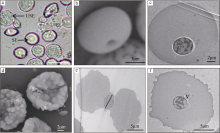
Fig. 1
The oocytes in the early growth stage of Siphonosoma australe. (a) smooth erythrocyte (SE) and uneven surface erythrocyte (USE); (b) and (c) red blood cell with a smooth surface (SEM/TEM); (d) disk-like cells with the uneven surfaces (SEM); (e) double-side concave oocyte with vesiculose, lumpy and corrugated surface (TEM); (f) oocyte with an uneven surface (TEM). V: vesicle"

Fig. 1
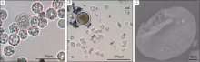
Fig. 2
The oocytes in the late growth stage of Siphonosoma australe. (a) 30μm oocyte; (b) oocyte beginning to form vitelline membrane; (c) 40μm cookie oocyte with the intracellular yolk granules (SEM)"

Fig. 2
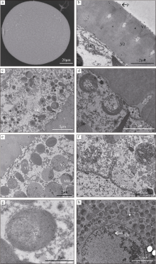
Fig. 3
The oocytes in the early mature stage of Siphonosoma australe. (a) spherical oocyte, membrane pores masked by a superficial vitelline membrane (SEM); (b) sunken membrane-pores and two layers (V1 and V2) forming the vitelline membrane; (c) ~ (e) morphology of a variety of yolk particles and numerous organelles (TEM); (f) numerous endoplasmic reticulum appearing around the nuclear membrane (TEM); (g) yolk particles surrounded by the Golgi (TEM); (h) chromatin forming clumps with high electron density (TEM). V1: outer layer of vitelline membrane; V2: inner layer of vitelline membrane; P: granular protuberance; MPV: micropinocytotic vesicle; L: lysosome; G: Golgi body; M: mitochondria; ER: endoplasmic reticulum; Y1: type I yolk granule; Y2: type II yolk granule; MB: myeloid body; Ch: chromatin"

Fig. 3
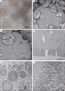
Fig. 4
The oocytes in the late mature stage of Siphonosoma australe. (a) mature oocytes (120 μm in diameter); (b) exfoliation of the outer-layer membrane (SEM); (c) clear visible micropores after peeling of outermost membrane; (d) micropinocytotic vesicles in the vitelline membrane; (e) large number of yolk particles, mainly contained by the Golgi bodies; (f) yolk granules filling to the cytoplasm of a mature oocyte. PM: magnified membrane-pore structure, Y2: type II yolk particles formed by Golgi wrapping, N: nucleus"

Fig. 4

Fig. 5
Monthly changes of composition and density of oocyte in the coelom of Siphonosoma australe"
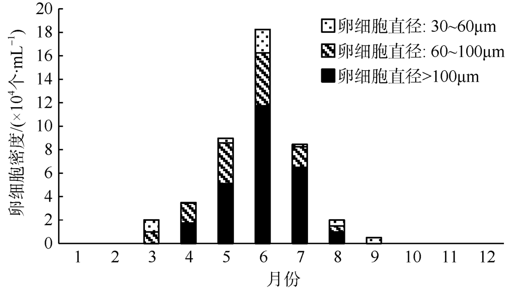
Fig. 5
| [1] | 陈慧, 林国文, 陈武, 等, 2009. 可口革囊星虫生殖周期的观察[J]. 海洋渔业, 31(2): 139-145. |
| CHEN HUI, LIN GUOWEN, CHEN WU, et al, 2009. Observation on the reproductive cycle of Phascolosoma esculenta[J]. Marine Fisheries, 31(2): 139-145. (in Chinese with English abstract) | |
| [2] | 陈子安, 杜晓东, 王庆恒, 等, 2007. 3种星虫线粒体16S rRNA、CO I和Cyt b基因片段的序列比较[J]. 广东海洋大学学报, 27(4): 3-10. |
| CHEN ZI’AN, DU XIAODONG, WANG QINGHENG, et al, 2007. Sequence comparison and phylogenetic analysis of mtDNA 16SrRNA and CO I and Cyt b gene fragments in three species of Sipuncula[J]. Journal of Guangdong Ocean University, 27(4): 3-10. (in Chinese with English abstract) | |
| [3] | 顾晓英, 竺俊全, 许式见, 等, 2009. 可口革囊星虫(Phascolosoma esculenta)卵子发生的组织学研究[J]. 海洋与湖沼, 40(3): 283-288. |
| GU XIAOYING, ZHU JUNQUAN, XU SHIJIAN, et al, 2009. Oogenesis of Phascolosoma esculenta: histological study[J]. Oceanologia et Limnologia Sinica, 40(3): 283-288. (in Chinese with English abstract) | |
| [4] | 郭学武, 李复雪, 1993. 光裸星虫生殖周期的研究[J]. 热带海洋学报, 12(2): 69-76. |
| GUO XUEWU, LI FUXUE, 1993. Studies on the reproductive cycle of Sipunculus nudus linnaeus[J]. Tropic Oceanology, 12(2): 69-76. (in Chinese with English abstract) | |
| [5] | 兰国宝, 阎冰, 2002. 方格星虫繁殖生物学研究[J]. 水产学报, 26(6): 503-509. |
| LAN GUOBAO, YAN BING, 2002. The reproductive biology of peanut worm, Sipunculus nudus[J]. Journal of Fisheries of China, 26(6): 503-509. (in Chinese with English abstract) | |
| [6] | 李凤鲁, 周红, 王玮, 1992. 中国沿海管体星虫属(星虫动物门)的研究[J]. 青岛海洋大学学报, 22(1): 97-102. |
| LI FENGLU, ZHOU HONG, WANG WEI, 1992. Studies on the genus Siphonosoma (sipuncula) off the China coasts[J]. Journal of Ocean University of Qingdao, 22(1): 97-102. (in Chinese with English abstract) | |
| [7] | 刘懂, 龙玲利, 曾海祥, 等, 2014. 可口革囊星虫体腔中卵细胞大小组成的周年变化[J]. 生物学杂志, 31(4): 29-32. |
| LIU DONG, LONG LINGLI, ZENG HAIXIANG, et al, 2014. Annual variation of egg size in the coelom of Phascolosoma esculenta[J]. Journal of Biology, 31(4): 29-32. (in Chinese with English abstract) | |
| [8] | 王庆恒, 杜晓东, 黄洪艳, 等, 2005. 湛江地区光裸星虫的生殖细胞发育和生殖周期[J]. 湛江海洋大学学报, 25(1): 5-9. |
| WANG QINGHENG, DU XIAODONG, HUANG HONGYAN, et al, 2005. Development of germ cells and reproductive cycle of Sipunculus nudus L in Zhanjiang[J]. Journal of Zhanjiang Ocean University, 25(1): 5-9. (in Chinese with English abstract) | |
| [9] | 王庆恒, 张家炜, 郝瑞娟, 等, 2017. 光裸星虫体腔液中卵子发生的超微结构[J]. 海洋与湖沼, 48(1): 57-66. |
| WANG QINGHENG, ZHANG JIAWEI, HAO RUIJUAN, et al, 2017. Ultrastructure of oogenesis in coelomic fluid of Sipunculus nudus[J]. Oceanologia et Limnologia Sinica, 48(1): 57-66. (in Chinese with English abstract) | |
| [10] | 吴斌, 1999. 光裸方格星虫(Sipunculus nudus L.)生殖细胞及胚胎发育[J]. 广西科学, 6(3): 222-226. |
| WU BIN, 1999. Development of genital cells and embryo of Sipunculus nudus L.[J]. Guangxi Sciences, 6(3): 222-226. (in Chinese with English abstract) | |
| [11] | 徐海娇, 2012. AFM研究高尔基体和内质网的结构[D]. 长春: 东北师范大学. |
| XU HAIJIAO, 2012. Studying the structure of Golgi complex and endoplasmic reticulum by AFM[D]. Changchun: Northeast Normal University. (in Chinese with English abstract) | |
| [12] | 周红, 李凤鲁, 王玮, 2007. 中国动物志无脊椎动物第四十六卷星虫动物门螠虫动物门[M]. 北京: 科学出版社. |
| ZHOU HONG, LI FENGLU, WANG WEI, 2007. Fauna sinica:invertebrata vol. 46. sipuncula echiura[M]. Beijing: Science Press. (in Chinese) | |
| [13] | 竺俊全, 王伟, 丁理法, 2012. 可口革囊星虫(Phascolosoma esculenta)卵黄合成期卵母细胞发育及卵黄发生与卵膜形成的超微结构[J]. 海洋与湖沼, 43(4): 870-876. |
| ZHU JUNQUAN, WANG WEI, DING LIFA, 2012. Ultrastructural observation on oogenesis, vitellogenesis and vitelline membrane formation during yolk-synthesizing stage in Phascolosoma esculenta[J]. Oceanologia et Limnologia Sinica, 43(4): 870-876. (in Chinese with English abstract) | |
| [14] |
AMOR A, 1993. Reproductive cycle of Golfingia margaritacea, a bipolar sipunculan, in subantarctic water[J]. Marine Biology, 117(3): 409-414.
doi: 10.1007/BF00349316 |
| [15] | ANDREWS E A, 1889. The reproductive organs of Phascolosoma gouldii[J]. Zoologischer Anzeiger, 12: 140-142. |
| [16] | CATALAN M A B, YAMAMOT M, 1994. Annual reproductive cycle of two Japanese species of sipunculans: Siphonosoma cumanense (Sipunculidae) and Phascolosoma scolops (Phascolosomatidae)[J]. Pacific Science, 48(2): 145-157. |
| [17] |
GONSE P H, 1956. L’ovogenese chez Phascolosoma vulgare: Ⅱ. Recherches biométriques sur les ovocytes[J]. Acta Zoologica, 37(3): 225-233.
doi: 10.1111/j.1463-6395.1956.tb00045.x |
| [18] | RICE M E, 1974. Gametogenesis in three species of sipuncula: Phascolosoma agassizii, Golfingia pugettensis, and Themiste pyroides[J]. La Cellule, 79(2-3): 259-313. |
| [19] | RICE M E, 1988. Observations on development and metamorphosis of Siphonosoma cumanense with comparative remarks on Sipunculus nudus (Sipuncula, Sipunculidae)[J]. Bulletin of Marine Science, 42(1): 1-15. |
| [20] | RICE M E, 1989. Comparative observations of gametes, fertilization, and maturation in sipunculans[C]// Proceedings of the 23rd European Marine Biology Symposium. Fredensborg: Olsen and Olsen: 167-183. |
| [21] | SAWADA N, 1975. An electron microscopic study on the oogenesis of Sipunculan worms[C]// Proceedings of International Symposium on the Biology of the Sipuncula and Echiura. Belgrade: Nauč-no Delo Press: 169-176. |
| [22] |
YING XUEPING, DAHMS H U, LIU XINMEI, et al, 2009. Development of germ cells and reproductive biology in the sipunculid Phascolosoma esculenta[J]. Aquaculture Research, 40(3): 305-314.
doi: 10.1111/j.1365-2109.2008.02093.x |
| [1] | ZHAO Zhongwei, WU Lingyun, GAO Weijian, LI Wei. A study of the effect of shore platform morphology on coastal erosion of rocky cliffs in the Wucaiwan Bay, E’man, Hainan Island [J]. Journal of Tropical Oceanography, 2024, 43(5): 106-115. |
| [2] | XU Bingjie, LIU Yiming, LIAN Changpeng, WU Tao, PAN Ying. Ovarian development, oocyte and yolk production of Paphia textile in Beibu Gulf, Guangxi [J]. Journal of Tropical Oceanography, 2024, 43(2): 48-58. |
| [3] | ZENG Weite, ZHANG Dongqiang, LIU Bing, YANG Yongpeng, ZHANG Hangfei, WU Duoyu, WANG Xiaolin. Distribution, main controlling factors and pollution assessment of heavy metals in surface seawater of the Northern Bay of Hainan Island, south China [J]. Journal of Tropical Oceanography, 2023, 42(6): 156-167. |
| [4] | WU Tao, PAN Ying, LIAN Changpeng, LIU Yiming, XU Bingjie, WANG Chaoqi, YANG Ling. The study on ovarian development, oogenesis and vitellogenesis of Lutraria sieboldii in Beibu Gulf of Guangxi [J]. Journal of Tropical Oceanography, 2023, 42(6): 137-149. |
| [5] | HUANG Peixian, YAO Xuemei, YU Qiaochi, ZHANG Jiayu. Mitogenome characteristics and phylogenetic analysis of Siphonosoma australe in Hainan [J]. Journal of Tropical Oceanography, 2023, 42(2): 54-63. |
| [6] | LIAN Changpeng, WU Tao, WANG Chaoqi, YANG Ling, PAN Ying. Ovarian development, histology of oogenesis and yolk formation of Tapes conspersus from the Beihai Yingpan, Guangxi [J]. Journal of Tropical Oceanography, 2022, 41(5): 170-179. |
| [7] | LIU Jinmei, HUANG Bingxin, DING Lanping, WANG Xuecong, YAN Jing, YAN Panzhu, ZHANG Yao. Morphological and phylogenetic studies of Yonagunia formosana in southeastern Hainan province, China [J]. Journal of Tropical Oceanography, 2021, 40(6): 76-82. |
| [8] | WANG Yuanqi, YANG Yang, ZHOU Liang, WANG Yaping, GAO Shu. Interpreting the origin of coastal boulders on a coral reef flat at Xiaodonghai of Hainan Island based on storm wave energy analysis [J]. Journal of Tropical Oceanography, 2021, 40(4): 110-121. |
| [9] | MENG Tian, CHEN Zuo, ZHU Jun, ZOU Xiaoxiao, FU Qingyan, BAO Shixiang. Morphological and molecular study on marine green alga, Halimeda velasquezii, first recorded in Hainan Island [J]. Journal of Tropical Oceanography, 2020, 39(6): 114-121. |
| [10] | Yongmei LI, Rui LIU, Nan YANG, Lanping DING, Bingxin HUANG, Yixiao WANG, Yanwei CHEN. Morphological taxonomy of four species of genus Gracilaria (Gracilariaceae, Rhodophyta) from Hainan Province, China [J]. Journal of Tropical Oceanography, 2018, 37(4): 29-37. |
| [11] | Xingru FENG, Jinyuan LI, Baoshu YIN, Dezhou YANG, Haiying CHEN, Guandong GAO. Characteristics of ocean waves in coastal area of Dongfang, Hainan Island based on observations [J]. Journal of Tropical Oceanography, 2018, 37(3): 1-8. |
| [12] | Junpeng ZHAO, Yihan LI, Wenping GONG. The seasonal and sporadic variation of the spreading of a small tropical river plume and its associated dynamics [J]. Journal of Tropical Oceanography, 2017, 36(4): 67-76. |
| [13] | Tiantian GUO, Shengbo CHEN, Tianqi LU. The inversion of multiple-phase SSTs based on the MODIS data: a case study on the southwest coastal waters of Hainan Island [J]. Journal of Tropical Oceanography, 2017, 36(1): 9-14. |
| [14] | CHEN Biao, WANG Jing, JING Zhiyou, LI Min. Seasonal and interannual variation of thermal fronts off the east coast of Hainan Island and the west coast of Guangdong [J]. Journal of Tropical Oceanography, 2016, 35(5): 1-9. |
| [15] | TIAN Yuhang, CHEN Zhong, LIU Jirui, HUANG Weixia, ZHONG Yi. Influence of the grain size on the porosity and acoustic velocity of offshore surface sediments in the Southeastern Hainan Island [J]. Journal of Tropical Oceanography, 2016, 35(3): 48-54. |
|
||